STRESS FRACTURES
STRESS FRACTURES. DAVE HAIGHT, MD Sports Medicine Fellow April 2009. Outline. Pathophysiology Risk Factors Associations Diagnosis General Treatment Specific Cases. CAUSE. Change in load Small number of repetitions with large load Large number of reps, usual load

STRESS FRACTURES
E N D
Presentation Transcript
STRESS FRACTURES DAVE HAIGHT, MD Sports Medicine Fellow April 2009
Outline • Pathophysiology • Risk Factors • Associations • Diagnosis • General Treatment • Specific Cases
CAUSE • Change in load • Small number of repetitions with large load • Large number of reps, usual load • Intermediate combination of increased load and repetition
PATHOPHYSIOLOGY • Wolff’s Law: change in external stress leads to change in shape and strength of bone • bone re-models in response to stress • ABRUPT Increase in duration, intensity, frequency without adequate rest (re-modeling) • Stress fracture: imbalance between bone resorption and formation • Microfracture -> continued load -> stress fracture
EPIDEMIOLOGY • 1% of general population • 1-8% of collegiate team sports • Up to 31% of military recruits • 13-52% of runners
RISK FACTORS • History of prior stress fracture • Low level of physical fitness, non-athlete • Increasing volume and intensity • Female Gender • Menstrual irregularity • Diet poor in calcium • Poor bone health • Poor biomechanics
RISK FACTORS cont • Prior stress fracture: • 6 x risk in distance runner and military recruits • 60% of track athletes have hx of prior stress fracture • One year recurrence: 13% • Poor Physical Fitness - muscles absorb impact • >1cm decrease in calf girth • Less lean mass in LE • Less than 7 months prior strength training
INTRINSIC FACTORS • Extreme arch morphologies: • Pes cavus • Pes planus • Biomechanical factors: • Shorter duration of foot pronation • Sub-talar joint control • Tibial striking torque • Early hindfoot eversion
EXTRINSIC FACTORS • Activity type and intensity • Footwear • Older shoes • Shock absorbing cushioned inserts • Running Surface • Treadmill • Track
Ballet: Runners: Sprinters: Long dist runner: Baseball, tennis: Gymnasts: Rowers, golfers: Hurdlers: Rowers, Aerobics: Bowling, running: Lumbar, femur, metatarsal Tibia, metatarsal Navicular Femoral neck, pelvis Humerus Spine, foot, pelvis Ribs Patella Sacrum Pelvis ASSOCIATIONS
Classic Clinical History • Change in training or equipment • Gradual onset over 2 to 4 weeks • Initially pain only with activity • Progresses to pain after activity • Eventually constant pain with ADLs
DIAGNOSISHistory • Sports participation • Significant change in training • Hills, surface, intensity • Dietary History: adequacy, Vit D, Calcium • Menstrual History • General Health • Occupation • Past medical history • Medications • Family history (osteoporosis)
IMAGINGX-ray • Only ~ 30% positive on initial examination • 10 - 20% never show up on plain films • If a positive x-ray • Localized periosteal reaction • Radiolucent line • Cancellous bone - band-like focal sclerosis
Bone Scan • 95% show up after 1 day • Extremely sensitive but not as specific with up to 24% false-positive results (stress reaction) • Differentiate between acute and old lesions • Acute stress fracture: three phase positive • Shin splint: delayed phase only
MRI vs. bone scan, CJSM 2002 • MRI less invasive, provided more information than bone scan and recommended for initial diagnosis and staging of stress injuries • “Limited” MRI may be cheaper than bone scan at some institutions
How I Decide Between an MRI and Bone Scan • MRI • Usually can be done more quickly (1 vs. 4 hours) and scheduled for a sooner date • No radiation • Better soft tissue detail • Bone Scan • Covers a wider area of the body (if bilateral or diffuse symptoms) • Sometimes easier to interpret • Cheaper
RADIATION COMPARISON Study mSvrelative radiation Plain film foot <0.01 < 1.5 days Plain film CXR 0.02 2.4 days Plain film pelvis 0.7 3.2 mo Tech-99 bone scan 3 (150 CXR) 1.2 yrs CT L-spine 6 (300 CXR) 2.3 yrs CT abd / pelvis 10 (500 CXR) 4.5 yrs
GENERAL TREATMENT • PROTECTION • Reduce pain • Promote healing • Prevent further bone damage • ADLs are permitted • Stretching and flexibility exercises • Cross-training (non-weight-bearing exercise) • Modified rest for six to eight weeks (or until pain-free for two to three weeks)
ACTIVITY MODIFICATION • Activity should be pain free • Approximate desired activity • Cycle • Swim • Walk • Elliptical • Deep water running • MUST BE PAIN FREE
REHAB EXERCISE • Address biomechanical issues: • Muscle inflexibility • Limb Length Discrepancy • Excessive pronation, pes cavus, pes planus • Replace running shoes • Strength training
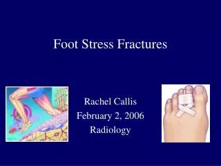
Site of Stress Fractures • Tibia - 39.5% • Metatarsals - 21.6% • Fibula - 12.2% • Navicular - 8.0% • Femur - 6.4% • Pelvis - 1.9%
High Risk Talus Tarsal navicular Proximal fifth metatarsal Great toe sesamoid Base of second metatarsal Medial malleolus High Risk Pars interarticularis Femoral head Femoral neck (tension side) Patella Anterior cortex of tibia (tension side) HIGH RISK
High-Risk Tibial Stress Fx • Anterior, middle-third stress fractures are very concerning • Tension side of bone • May present like shin splints • Seen more commonly in jumpers and leapers • See “dreaded black line” on x-ray • Heal very poorly
Dreaded Black Line
Management of High-Risk Tibial Stress Fx • Immobilization • 4-6 months of rest • Pulsed low-intensity U/S or electrical stimulation may decrease symptoms and speed return to activity, 30 minutes/day x 3-9 mos. • IM rod for failed conservative or patient preference
5th metatarsal stress fracture
Mgmt. of 5thMetatarsal Stress Fx • High risk for delayed union or nonunion • Non-weight-bearing cast for 6 weeks versus IM screw fixation
Femoral Stress Fx • Primary presenting symptom is groin pain; possibly thigh or knee pain • Hip motion may be painful • Hop test • Fulcrum test for shaft fx
Femoral Neck Stress Fx • Early diagnosis critical • If x-rays negative, bone scan/MRI • MRI diagnostic imaging of choice for femoral neck stress fractures
Femoral Neck Stress Fx • Compression side. • Inferior part of femoral neck • Younger patients • Less likely to become displaced • Complications possible • Treatment-non-weight bearing, followed by touch-down WB, then partial WB over a total of 8-12 weeks
Femoral Neck Stress Fx • Distraction side • Superior cortex or tension side of neck • High propensity to become displaced • Frequent complications • Treated acutely with internal fixation
Tarsal Navicular Stress Fx • Consider in: Sprinters, Jumpers, Hurdlers, Basketball, Football • Mean interval of 7 -12 months before diagnosis • Vague mid-foot pain • Pain on dorsum of foot • Foot cramping
Tarsal Navicular Stress Fx • X-rays usually negative • Bone scan vs. MRI vs. thin cut CT
Mgmt. of Navicular Stress Fx • Most studies suggest that allowance of weight-bearing immediately after diagnosis increases the non-union rate • General/simple rules: • (+) bone scan/MRI/CT and/or incomplete fx- NWB cast x 6-8 weeks, and then gradual rehab • Complete fx and/or bony sclerosis- ORIF with compression screw +/- bone graft
Orthopedic Consultation • High Risk Fracture sites • Femoral Neck - tension side • Navicular • 5th Metatarsal • Anterior tibial shaft • High Level Athlete/Laborer • Failed conservative therapy
PREVENTION • Small incremental increases in training FITT • Shock absorbing shoe/boot inserts • Calcium 200mg, Vit D 800IU (27% decr.) • OCPs: sig increase in bone mineral density, no impact on stress fracture rate • Modification of female recruit training: • Lower march speed • Softer surface • Individual step length/speed • Interval training instead of longer runs
Take Home Points • Avoid a delay in diagnosis, image early • REST is a 4-letter word to athletes; thus advise relative rest, allowing for cross-training or unaffected body-training during the healing period
Take Home Points • Correct underlying nutritional, hormonal or biomechanical abnormalities to promote healing and prevent recurrence • Despite our best efforts, some athletes will never return to their pre-injury level of competition due to some specific stress fractures (navicular, femoral neck, anterior tibia)