Download

1 / 20
210 likes | 485 Vues
Stress Fractures. (emphasis on Medial Tibial ). Stress Fracture-Scope of Disease. First described by Breithraupt in 1855 5 th metatarsal in marching Prussian soldiers (march fx ) Incidence around 10% of all musculoskeltal injuries (including military training)

E N D
Stress Fractures (emphasis on Medial Tibial)
Stress Fracture-Scope of Disease • First described by Breithraupt in 1855 • 5th metatarsal in marching Prussian soldiers(march fx) • Incidence around 10% of all musculoskeltal injuries (including military training) • 95% of all stress fractures occur in lower extremity • 46% tibia • 15% navicular • 12% the fibula
Pathophysiology • Repetitive loading alters bone’s microstructure (↑ number & size microfx) • Response is ↑’d oseteoclastic & osteoblastic activity • Usually results in a stronger bone able to withstand greater loads • Initially osteoblasticactivity lags behind resorptive properties of osteoclasts • Process leaves bone susceptible to fatigue failure if the area is continually stressed without adequate time for repair • Couple this w muscle dysfxnfrom overuse results in focal bending stresses exceeding structural & physiologic tolerance of bone Usually takes at least 2-3 weeks to develop
Extrinsic Factors • Training Regime Too Much Too Hard Too Soon (10% Rule) • Equipment • Shoes • Right Shoe (pronator vs. supinator) • Old Shoe (change shoes every 500 miles-6 months) • Running Surface • Nutritional Habits • Calcium/Vit D (1000mg/800IU) • Adequate Calories • Rest • Medications- DepoProvera (? other progestins)
Intrinsic Factors • Muscle Endurance (most outshape, highest risk) • Excessive Hip External Rotation >60 deg • Leg Length Abnormality • PesCavus • Morton’s Foot hypermobile 1st ray &long 2nd ray • Hormonal Factors (lack of estrogen- ♀ athlete triad)
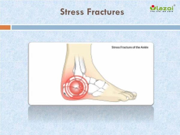
Diagnosis • First Pain at end of training • Then during whole training session • Then continuous • Pain w palpation • Swelling • Fulcrum Test • Hop Test Definitive Dx requires radiographic confirmation
Imaging • Plain Films-findings lag behind or nonexsistent • Bone Scan Sens 84-100%- poor specf-can’t use for f/u • MRI-high sens and spec and good for f/u • CT-excellent bone detail but ↑ radiation • U/S-operator and location dependent Sensitivity % of pts w disease who test + Specificity % of pts w/o disease who test -
(A) Endosteal remodeling and cortical thickening (arrow) consistent with a stress fracture. (B) Sagittal high-resolution fast spin echo MRI of same patient demonstrating moderate cortical thickening and a faint cortical fracture line (arrow).
phase 1 (flow phase) phase 2 (blood-pool phase) phase 3 (delayed phase) Soft tissue injuries increased uptake in only phases 1 and 2 Shin splints have increased signal only in the delayed-phase images Stress Fractures are + in all phases but take months to years to go to normal Must correlate with symptoms
(A) Sagittal fast inversion recovery MRI of lateral ankle margin with bone marrow edema pattern in the fibula with periosteal reaction consistent with stress fracture. (B) Sagittal fast spin echo MRI in the same patient demonstrates mild ill-defined endosteum and periosteal new bone formation (arrow ).
Sagittal T2 fat saturation showing diffuse marrow edema. Sagittalshowing diffuse marrow edema.
Ultrasound showing periosteal elevation and increased blood flow
Grading Scale Time to Heal 3.3 Weeks 5.5 Weeks 11.4 Weeks 14.3 Weeks Arendt EA, Griffiths HJ. The use of MR imaging in the assessment and clinical management of stress reactions of bone in high-performance athletes. Clin Sports Med 1997;16:291–306
Differential Diagnosis M M MTSS Stress Fx M ECS • Muscle strain • Medial/Anterior tibia stress syndrome (shin splints-MTSS/ATSS) • Stress reaction • Exertional Compartment Syndrome (ECS) • Infection • Bursitis • Neoplasm • Nerve entrapment MTSS Stress Reaction Stress Fracture Continuum
Treatment • Relative Rest and Activity Modification • Pneumatic Braces • Foot Orthoses • Pulsed U/S (little less force than lithotripsy) • Bone Stimulators • Bisphosphonate (shuts down osteoclasts) • ? Avoid NSAIDs (Cox2 is needed for fx repair) • Correct Training Errors (no more 10% ↑ week) • Improve Muscular and Aerobic Capacity
Anterior Tibial Cortex “Dreaded Black Line”
Stress Fx vs. Insufficiency Fx Abnl Stress to Nl bone Nl stress to Abnl Bone Cortical Fx vs. CancellousFx(consider DEXA if not good hx) Long Bones Femoral Neck/Talus/Navicular Low-grade fx at a low-risk site can be allowed to continue to compete Low-grade fx at a high-risk site needs to heal before full return to activity High Risk Femoral Neck Patella Anterior tibialdiaphysis Talus TarasalNavicular 5th Metatarsal Low Risk Femoral shaft Medial tibia Ribs Ulna shaft 1st -4th Metatarsals