1 / 0

Stress Fractures

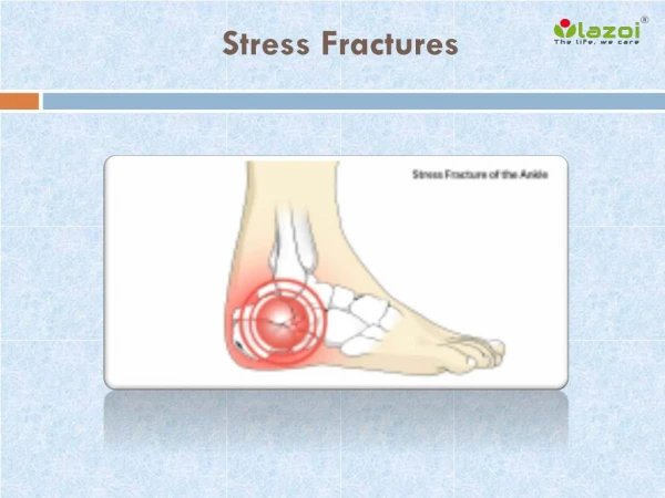
Stress Fractures. Kevin deWeber, MD, FAAFP, FACSM Director, Military Sports Medicine Fellowship USUHS/Ft. Belvoir 2011 (many slides courtesy Dave Haight , MD. Outline. Pathophysiology Risk Factors Associations Diagnosis General Treatment Treatment of High-Risk Cases.

finna
Télécharger la présentation

Stress Fractures

An Image/Link below is provided (as is) to download presentation Download Policy: Content on the Website is provided to you AS IS for your information and personal use and may not be sold / licensed / shared on other websites without getting consent from its author. Content is provided to you AS IS for your information and personal use only. Download presentation by click this link. While downloading, if for some reason you are not able to download a presentation, the publisher may have deleted the file from their server. During download, if you can't get a presentation, the file might be deleted by the publisher.

E N D

Presentation Transcript


  1. Stress Fractures

    Kevin deWeber, MD, FAAFP, FACSM Director, Military Sports Medicine Fellowship USUHS/Ft. Belvoir 2011 (many slides courtesy Dave Haight, MD
  2. Outline Pathophysiology Risk Factors Associations Diagnosis General Treatment Treatment of High-Risk Cases
  3. Stress fracture [stress frack-chur] Break in a normal bone after it is subjected to repeated tensile or compressive stresses, none of which would be large enough individually to cause the bone to fail, in a person who is not known to have an underlying disease that would be expected to cause abnormal bone fragility.
  4. Insufficiency Fracture[in-suh-fish-n-see frack-chur] Fracture due to repeated compressive or tensile stresses in a bone whose mechanical strength is reduced due to a condition that is present either throughout the skeleton (osteoporosis, osteomalacia, osteogenesisimperfecta, etc.) or in a bony region (eg, demineralization in a limb due to disuse).
  5. Pathologic fracture[path-o-lah-jickfrack-chur] Fracture due to a localized loss of strength in a bone from a disease process immediately underlying the bone, eg, bone tumors , bone cysts, and infections.
  6. PREVALENCE 1% of general population get ‘em 1-8% of collegiate team athletes get ‘em Up to 31% of military recruits get ‘em 13-52% of runners get ‘em
  7. Most Common Sites of Stress Fractures Tibia - 39.5% Metatarsals - 21.6% Fibula - 12.2% Navicular - 8.0% Femur - 6.4% Pelvis - 1.9% OTHER – 10.4% Weight- bearing
  8. CAUSE Change in load(force on the bone) Small number of repetitions with large load Large number of reps, usual load Intermediate combination of increased load and repetition
  9. PATHOPHYSIOLOGY Stress fracture: imbalance between bone resorption and formation Wolff’s Law: change in external stress leads to change in shape and strength of bone bone re-models in response to stress ABRUPT increase in duration, intensity, frequency without adequate rest (re-modeling) Microfracture -> continued load -> stress fracture
  10. Review of Risk Factor Types Intrinsic: Gender, genetics Anatomical malalignment/ biomechanical Dietary Muscle weakness/imbalance Extrinsic Training errors Equipment mismatch Technique errors Environmental Sports-imposed deficiencies
  11. INTRINSIC RISK FACTORSfor Stress Fractures History of prior stress fracture Low level of physical fitness Female Gender Menstrual irregularity Diet poor in calcium and dairy Poor bone health Poor biomechanics
  12. INTRINSIC RISK FACTORS cont Prior stress fracture: 6 x risk in distance runner and military recruits 60% of track athletes have hx of prior stress fracture One year recurrence: 13% Poor Physical Fitness – [muscles absorb impact] >1 cm decrease in calf girth Less lean mass in LE < 7 months prior strength tng
  13. INTRINSIC RISK FACTORS cont Why female gender? [1.2-10x] Higher rates of other risk factors Poorer bone health, Menstrual irregularity, disordered eating Poor bone health: Supplementing female military recruits with Ca and Vit D reduced stress fracture incidence Lappe J, Cullen D, Haynatzki G, et al.. J Bone Miner Res 2008 FamHx osteoporosis: 3x risk
  14. INTRINSIC RISK FACTORS cont BIOMECHANICAL FACTORS Shorter duration of foot pronation Sub-talar joint control Tibial striking torque Early hindfooteversion Pescavus (unproven) Pesplanus (unproven) Tibial stress Fractures (retrospective)
  15. EXTRINSIC FACTORS Increasing volume and intensity Footwear Older shoes Absence of shock absorbing inserts Running Surface?: mixed results Treadmill vs Track Activity type
  16. ACTIVITY TYPE ASSOCIATIONS Ballet: spine, femur, metatarsal Runners: tibia, MT Sprinters: navicular Long dist runner: femoral neck, pelvis Baseball, tennis: humerus Gymnasts: spine, foot, pelvis Rowers, golfers: ribs Hurdlers: patella Rowers, Aerobics: sacrum Bowling: pelvis
  17. Classic Clinical History Gradual onset of pain over weeks to months Initially pain only with activity “can’t run through it” Progresses to pain after activity Eventually constant pain with ADLs Change in training regimen “too much too soon” Change in equipment Shoes, etc.
  18. DIAGNOSIS: take a good History Sports participation Occupation Significant change in training Hills, surface, intensity Dietary History: adequacy, Vit D, Calcium Menstrual History General Health Past medical history Medications Family history (osteoporosis)
  19. DIAGNOSIS: Examination “High-Speed Digital Imaging” Localized tenderness to palpation in a location known to commonly sustain stress fractures Fulcrum test? Neither sensitive nor specific Hop test? Neither sensitive nor specific Risky in high risk fractures Tuning fork? 43% sensitive, 49% specific
  20. DIAGNOSIS: Biomechanical Evaluation Leg length discrepancy 70% incidence in patients w/ LE stress fractures Joint range of motion and ligamentous stability Muscle strength and flexibility Limb alignment (eg, genuvarus or valgus) Foot type (eg, pescavus or planus) Gait analysis in shoes worn during physical activity Core muscle strength (eg, abdominal, back, and hip musculature)
  21. IMAGING: X-ray Poor sensitivity ~ 30% positive on initial examination 4 Possible findings Localized periostealelevation Cortical thickening Focal sclerosis Radiolucent line 10 - 20% never show up on plain films
  22. Early Metatarsal Stress Fracture
  23. One Week Later…..
  24. Imaging: Bone Scan Extremely sensitive 95% show up after 1 day Not very specific up to 24% false-positive results (stress reaction) Differentiate between acute and old lesions Acute stress fracture: all three phases positive Shin splint: delayed phase only
  25. Imaging: CT scan Excellent fracture line detail More specific than bone scan Useful for fracture age/non-union Pars interarticularis, sesamoids, etc. DOWNSIDE: high radiation dose
  26. Imaging: MRI Highly sensitive (= bone scan) More specific than bone scan still not perfect Cost may be lower than bone scan some places Non-invasive, no radiation Sensitive for soft tissue injury DOWN: less cortical bony detail than CT
  27. Imaging: Ultrasound Useful if fracture superficial Shows hematoma, hypervascularity, periosteal elevation, cortical defect Metatarsal fractures: sensitivity 83%, specificity 76% Balal F, Gandjbakhch F, Foltz V et al. J Rheumatol 2009
  28. MRI vs. bone scan, CJSM 2002 MRI less invasive, provided more information than bone scan and recommended for initial diagnosis and staging of stress injuries “Limited” MRI may be cheaper than bone scan at some institutions
  29. RADIATION COMPARISON Study mSvrelative radiation Plain film foot <0.01 < 1.5 days Plain film CXR 0.02 2.4 days Plain film pelvis 0.7 3.2 mo Tech-99 bone scan 3 (150 CXR)1.2 yrs CT L-spine 6 (300 CXR) 2.3 yrs CT abd / pelvis 10 (500 CXR) 4.5 yrs
  30. Differential Diagnosis Muscle strains Tendinopathy Nerve entrapment Medial tibial stress syndrome Neoplasm Infection
  31. HIGH RISK STRESS FRACTURES Talus Tarsal navicular Proximal fifth metatarsal Great toe sesamoid Base of second metatarsal Medial malleolus Pars interarticularis Femoral head Femoral neck (tension side) Patella Anterior cortex of tibia (tension side)
  32. GENERAL TREATMENT for LOW-RISK STRESS FRACTURES PROTECTION Reduce pain Promote healing Prevent further bone damage ACTIVITY MODIFICATION Rest from painful activities 6-8 weeks (or until pain-free for two to three weeks) Cross-training (non-painful exercise) REHABILITATIVE EXERCISE Flexibility, strength balance BIOMECHANICAL CORRECTIONS
  33. Early Treatment Speeds Recovery! Evaluation < 3 weeks of sx onset: 10.4 weeks to RTP Evaluation > 3 weeks 18.4 weeks to RPT Ohta-Fukushima M, Mutoh Y, Takasugi S, et al. J Sports Med Phys Fitness 2002
  34. ACTIVITY MODIFICATION Activity should be pain free Approximate desired activity Cycle Swim Walk Elliptical Deep water running
  35. REHAB EXERCISE and BIOMECHANICAL CORRECTIONS Muscle flexibility Strength training Excessive pronation, pescavus, pesplanus Limb Length Discrepancy Replace running shoes every… <300 miles
  36. Other Treatment Modalities Ultrasound: unsure 1 study no benefit, 2 studies + benefit ECSWT: maybe—consider in high-risk sites Worked in 1 small retrospective study Medications: Iloprost: 1 small retrospective study in subchondral stress fractures of knee Capicitatively Coupled Electric Fields: No benefit in recent RCT
  37. HIGH RISK STRESS FRACTURES High risk for delayed union , nonunion, refracture Talus Tarsal navicular Proximal fifth metatarsal Great toe sesamoid Base of second metatarsal Medial malleolus Pars interarticularis Femoral head Femoral neck (tension side) Patella Anterior cortex of tibia (tension side)
  38. High-Risk Tibial Stress Fracture Anterior, middle-third stress fractures are very concerning Tension side of bone May present like shin splints Seen more commonly in jumpers and leapers If you see “dreaded black line” on x-ray, poor prognosis
  39. Management of High-Risk Tibial Stress Fx 4-6 months of rest +/- immobilization ? Therapeutic US or SWT Surgery if not healing in 4-6 months Intramedullary rod
  40. Proximal 5th metatarsal stress fracture
  41. Mgmt. of 5th Metatarsal Stress Fracture Sxs <3 wks, neg x-rays: Avoid WB activity; 5th MT unloader orthotic Sxs > 3 wks or + x-rays Cast, NWB x 6 wks OR Screw fixation (faster RTP) Non-union: Screw-it!
  42. Lumbar Spondylolysis Stress fracture of the pars interarticularis Caused by repetitive hyper-extension Often develops in the teenage or pre-teen years May be bilateral
  43. Sports Associated with Spondy Football (offensive lineman) Gymnastics Wrestling Diving Tennis Volleyball
  44. Physical Exam- Spondy Tenderness to palpation over spines > paraspinal muscles Extension exacerbates pain “Stork test”—not very sensitive Tight hamstrings- cause or effect?
  45. Imaging Suspected Spondy AP & lateral x-rays r/o other bony causes Obliques don’t change management Also order SPECT bone scan MRI not as sensitive CT with fine cuts: prognostic Acute vs sclerotic
  46. Treatment- Spondy Relative rest from painful activity Physical Therapy referral Williams flexion-based exercises: Spine Surgeon referral Anti-lordotic bracing Brace 6 weeks - 6 months (controversial) Return to activity in brace when pain-free Surgery if fail treatment
  47. Femoral Neck Stress Fracture Vague anterior or medial groin/hip pain Early diagnosis critical Anterior hip tenderness Log-roll pain Pain with straight-leg-raise If x-rays negative, order MRI Crutches/NWB until ruled out! MRI diagnostic imaging of choice for femoral neck stress fractures
  48. Femoral Neck Palpation Iliopsoas bursa
  49. Femoral Neck Stress Fractures Compression side. Inferior part of femoral neck Less likely to become displaced Complications possible Treatment: Fatigue line <50% neck width: Crutches/NWB until asymptomatic, then relative rest 4-6 wks Fatigue line >50% neck width: surgical fixation
  50. Femoral Neck Stress Fx Tension side: HIGH-RISK Superior cortex or tension side of neck High propensity to become displaced Frequent complications Treated acutely with internal fixation
  51. Tarsal Navicular Stress Fx: HIGH-RISK Consider in: Sprinters, Jumpers, Hurdlers, Basketball, Football Mean interval of 7 -12 months before diagnosis “DON’T BE THAT GUY” WHO MISSES IT Vague mid-foot medial arch pain Foot cramping
  52. Tarsal Navicular Stress Fx X-rays usually negative MRI or thin-cut CT better than bone scan
  53. Tarsal Navicular Stress Fracture Meta-analysis 2010: NWB cast better than WB 6-8 weeks Semirigid orthotic during subsequent ambulation NWB trend better than surgery Torg JS, Moyer J, Gaughan JP, Boden BP. Am J Sports Med 2010. Nonunion/displacement: surgery
  54. Navicular Stress Fx Return to Play After casting, if no tenderness at the “N” spot, then can gradually return Reassess every 1-2 weeks, gradual return at 6 weeks if no symptoms AFTER 6 weeks of protection, 6 weeks of PT for strength and flexibility prior to return to run! Average return to play is 4-6 months Follow up radiography not helpful for return to activity
  55. Sesamoid Stress Fracture Risk: Sudden start-stop sports Repetitive forced dorsiflexion
  56. Sesamoid Stress Fracture Tx: NWB x 6 weeks with cast to tip of great toe to prevent DF Failure: Surgery (excision or grafting)
  57. Talus Stress Fracture Chronic pain after ankle sprain Location of fx: body near lateral process MRI or CT for best imaging Tx: 6-8 wks NWB in cast Failure to heal: Excision of lateral process
  58. Patella Stress Fracture Risk factors: cerebral palsy, hurdlers ACL reconstruction w/ BTB Tx if x-rays neg: Non-painful activity Tx if x-rays + or high-demand athlete K-wire fixation
  59. Femoral Head Stress Fracture Tx: NWB Ortho referral; high rate of arthroplasty
  60. Medial Malleolus Stress Fracture Seen in runners and jumpers Risk of non-union Tx if x-rays neg and MRI w/o fx line: SLWC or ankle brace; takes 4-5 MONTHS High-level athlete: surgery (FASTER RTP) Tx if x-rays + or non-union: Surgery
  61. Orthopedic Consultation High Risk Fracture sites High Level Athlete/Laborer Failed conservative therapy
  62. PREVENTION of STRESS FRACTURES Small incremental increases in training FITT Shock absorbing shoe/boot inserts Calcium 2000mg, Vit D 800 IU (27% decr.) Increased dairy products 62% decreased risk SF for each cup of skim milk Modification of female recruit training: Lower march speed Softer surface Individual step length/speed Interval training instead of longer runs ??: OCPs (sig increase in bone mineral density, no impact on stress fracture rate) NO: HCP selection of military recruits’ running shoes based on foot morphology 3 prospective studies by Knapik et al
  63. QUESTIONS?
More Related