Procedures Performed Outside the Operating Room by Dr. Masood Entezariasl
540 likes | 654 Vues
Learn about performing medical procedures in remote locations, including anesthesia services, equipment requirements, and managing anesthesia in invasive x-ray procedures like interventional radiology.

Procedures Performed Outside the Operating Room by Dr. Masood Entezariasl
E N D
Presentation Transcript
PROCEDURES PERFORMEDOUTSIDE THE OPERATING ROOM Dr Masood Entezariasl
Introduction • The number of diagnostic and therapeutic medical procedures that require specialized environments such that they must be performed away from traditional hospital and outpatient operating room suites is increasing (Table) • Office based anesthesia has become a primary mode of practice for many anesthesiologists
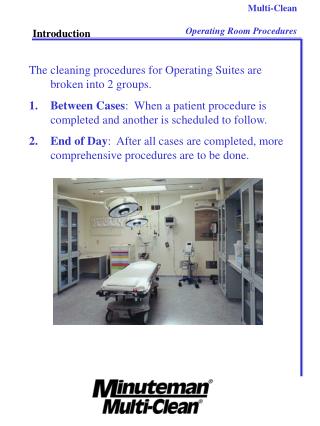
CHARACTERISTICS OF REMOTE LOCATIONS • Remote locations are much different from self-contained operating rooms • There should also be clear policies for dealing with remote equipment problems and unexpected escalations of medical problems • It is important for an anesthesiologist working in an unfamiliar remote location to keep track of the identity and role of personnel who participate in the surgical procedure or patient care • During times when the anesthesiologist may need experienced medical assistance (tracheal intubation, placement of a central venous catheter), it can be important to know which of the available staff members are qualifiedto render assistance • Readily availablepreoperative documentsfor all patients in remote locations must includethe attending surgeon's history and physical examination • Arrangements for patient arrival and check-in should be similar to those for outpatients and inpatients undergoing procedures in a traditional operating room setting
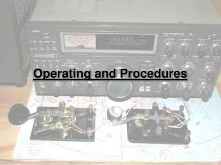
Remote locations must provide for the same basic anesthesia carethat is possible in any operating room • There must be adequate monitoring capabilities, the means to deliver supplemental oxygen by facemask with positive pressure, the availability of suction, equipment for providing controlled mechanical ventilation, an adequate supply of anesthetic drugs and ancillary equipment, and supplemental lightingfor procedures that involve darkness • Although new, portable anesthesia machines (Fig) can sometimes be placed very close to the patient to facilitate gas connections, it is often not possible to have an anesthesia machine as close to the patient as in the operating room (Fig) • The use of sedation, as for placement of a nerve block, should take place in an area (block room) where adequate equipment, drugs, and support personnel are available for immediate intervention • If anesthetic gases are to be used, scavenging must be sufficient to ensure that trace amounts are below the upper limits set by the Occupational Safety and Health Administration (OSHA)
Remote locations frequently involve additional hazards, such as exposure to radiation, high sound levels, and heavy mechanical equipment • Advance preparation should be made to have all needed equipment available, such as lead aprons, portable leadglass shields, and earplugs • At the end of the procedure one must often travel distances that are typically greater than the usual distance to the postanesthesia care unit or other patient units • So that patients can be safely and expeditiously taken to a recovery area, remote locations should always have available sufficient supplies of supplemental oxygen, appropriate transport monitoring equipment, and elevator and passageway keys • The anesthesiologist should always know the location of the nearest defibrillator, fire extinguisher, gas shutoff valves, and exits
Interventional Radiology -Neuroangiography and Body Angiography • Interventional neuroradiology (endovascular neurosurgery) mixes traditional neurosurgery with neuroradiology while also including certain aspects of head and neck surgery • Body angiography mixes general surgery with general radiology • In angiographic procedures the relevant blood vessel trees are imaged, after which a decision is made to continue by providing one or more therapeutic interventions via drugs or devices (or both)
MANAGEMENTOF ANESTHESIA • Anesthesia-related concernsinclude: • (1) maintenance of patient immobility and physiologicstability • (2) perioperative management of anticoagulation • (3)readiness for sudden unexpected complicationsduring the procedure • (4) provision of smooth and rapid emergence from anesthesiaand sedation at appropriate times (maybe required during the procedure) • (5) appropriate monitoring and management during transportafter completion of the procedure, particularly for critically ill patients, who may require continuous evaluationof breathing and systemic blood pressure
Physiologic Stability • Maintenance of blood pressure is particularly important in patients with cerebrovascular disease • Blood pressure targets should always be discussed preoperatively with the surgical team • Maintaining a higher than normal blood pressure is important in cases in which the patient has occlusive cerebrovascular disease, suchcases include patients undergoing emergency thrombolysis and patients with aneurysmal subarachnoid hemorrhage in whom vasospasm has developed • Conversely, prevention of blood pressure increases may be critical in certain groups , examples include patients with recently ruptured intracranial aneurysms or recently obliterated intracranial A-V malformation and patients who have undergone cerebrovascular angioplasty and stent placement in extracranial conductance vessels such as the carotid artery • These patients are also susceptible to post-treatment cerebral hyperperfusion injury and require careful control of systemic blood pressure after the procedure
Anticoagulation • Anticoagulation is often needed during intracranial catheter navigation to prevent thromboembolic complications • Heparin (70 U/kg) is commonly used to prolongthe baseline activated clotting time (ACT) by a factor of 2 to 3 • Hourly monitoring of the ACT is performed to assess the need for additional heparin, which can be given continuously or as intermittentboluses • Hemorrhagiccomplications during the procedure may necessitateemergency reversal of anticoagulation • If heparin has been administered, a full reversal dose of protamine (1 mg for each 100 units of heparin activity) should always be availablefor immediate injection • At the completion of uneventful procedures, heparin can be reversed with protamine, if deemed appropriate • Antiplatelet agents (aspirin, ticlopidine, and antagonists to glycoprotein IIb/IIIareceptors) are often used together with heparin, particularly when placing intraarterialstents • Although antiplatelet drugs decreasethe incidence of serious thromboembolic complications, emergency reversal of their anticoagulant effects is difficult • The only practical approach to antagonism of these drugs is empirical provision of exogenous platelets
Patient Transport • During transport from the imaging suite, airway management equipment, including a face mask and an Ambu bag or a Jackson-Rees circuit, should be immediately available for providing positive-pressure ventilation • It is not unusual to maintain an intravenous sedation regimen (propofol infusion) during transport or to have given additional medications just before transport
RADIATION THERAPY • Patient immobilityduring radiation therapy is the primary goal of sedation or general anesthesia so that the delivered radiation can be precisely targeted • Radiationtherapy may involve daily treatments for several weeks • Treatmentsfrequently take very little time, and patients want to quickly resume normal daily activities • In such instances, sedation or general anesthesia should be achieved with fast-onset, short-acting drugs appropriate for brief duration and rapid emergence while keeping in mind that sedation or anesthesia will be repeated daily • Anesthesia may also be required for lengthy or complex cases
Devices to Deliver Large Targeted Doses of Radiation • Radiation therapy for cancer delivers large radiation doses to target tissues • In some patients, radiation is used to kill vulnerable cancer cells while only minimally injuring noncancer cells • In others, radiation is used to kill all cells in the target region (reason that the radiation device may be referred to as a "knife") • Such radiation therapy is known as stereotactic because three-dimensional MRI and CT images are used by the radiation instrument to target specific tissue volumes. • GammaKnife simultaneously directs multiple carefully aligned, pencilthin gamma ray beams into the targeted area • TheCyberKnifeis also characterized by delivery of a large number of ovelapping pencil-thin gamma-ray beams to provide lethal radiation • In contrast to the GammaKnife, which exposes the patient to the gamma rays as a simultaneous single dose, the CyberKnifeexposes the patient to a sequence of several hundred gamma-ray beams, each being delivered from a computer-controlled robot arm that moves around the patient and shoots the beams at cancer regions from different directions
ANESTHESIA REQUIREMENTS • Anesthesia for GammaKnife procedures can involve general anesthesia or sedation for placement of a head frame, subsequent MRI or CT (or both), transport to the recovery room where anesthesia or sedation is maintained as one waits for generation of the computer data needed by the GammaKnife, and finally transport of the anesthetized or sedated patient to the GammaKnife room for the treatment • CyberKnife procedures require prior surgical implantation of radiopaque markers • The anesthesiologist must keep the anesthesia machine, the drug cart, and all tubes and hoses away from all locations that will be occupied by the robot arm • Robot motions can be monitored closely by a remote video connection to verify that one's setup is appropriate. • Scattered gamma radiation during therapy reaches high enough levels in the treatment room to requirethat all health care personnel be outside • Treatments occur in a heavily shielded room with health care personnel typically waiting on the other side of a large lead or iron door that takes 30 to 60 seconds to open • Physiologic monitoringis accomplishedvia two or more remote video connections
Alternative Radiation Therapy Delivery Devices • Other external beam radiation therapy modalities for cancer includeelectron beam radiation and heavy particle ion beam radiation • The "heavy particles" are nuclei from atoms larger than helium • This treatment mode has limited availability, but it has significant advantages because the energy deposition of a heavy ion beam is very concentratedand it can be targeted with millimeter precision • Intraoperative use of particle beams has become popular in cancer surgery • During intraoperative radiation therapy (IORT), a giant linear accelerator is placed in the operating room, and the depth and width of the electron beam are adjustedaccording to the patient's needs • Adjacent organs and tissues are shielded with lead • All personnel must leave the room during the radiation treatment, but thick shielding walls are not Necessary • A single IORT session will typically provide as much therapy as 10 to 20 daily gamma-raytreatments • In some instances the patient cannot receive IORT in the operating room when an intraoperative gamma-ray treatment is needed. In such cases, the anesthesiologist must transport the anesthetized patient to the alternative treatment area
ELECTROCONVULSIVE THERAPY • Electroconvulsive therapy (ECT) is used primarily after failure of pharmacotherapy for affective disorders, most notably severe depression, but also bipolar syndrome and schizophrenia • Because the ECT effect is evident within only a few treatments, it has been proposed for the treatment of psychiatric disorders of high acuity, such as suicidal patients or those unable to take food • There is little doubt about the short-term effectiveness of ECT for depression, but controversies remain regarding its place in long-term management, as well as the definition of failed pharmacotherapy. • ECT's therapeutic effects are thought to result from the release of neurotransmitters during the electrically induced grand mal seizure or, perhaps, from the reestablishment of neurotransmitter levels that occurs after seizure activity
Characteristics of Electrically Induced Seizures • It is important that an electrically induced seizure be of sufficient duration (>20 seconds) for optimal therapeutic effect • In this regard, the anesthesiologist must consider the impact of selected anesthetic drugs on the duration of seizure activity • Electrically induced seizures are characterized by an initial tonic phase (lasts 10 to 15 seconds), followed by a second myoclonic phase (lasts 30 to 60 seconds) • Seizure duration is monitored by motor activity and usually a single-channel electroencephalogram • Needs for adjustments in seizure length should be discussed with the anesthesiologist before ECT
Management of Anesthesia • The two goals of anesthetic management are to: (1) provide partial neuromuscular blockade because unmitigated motor activity can result in long bone fractures and skeletal muscle injury (2) render the patient briefly unconscious for applicationof the electrical stimulus • Theseizureis associated with a profound amnestic response and is not painful if motor activity is blocked • In the past, anesthetics most often used for ECT were the short-acting barbiturates methohexital (0.5 to 1.0 mg/kg IV) and thiopental(1.0-2.0 mg/kg IV) • More recently, propofol (1mg/kg IV) and etomidate(0.3 mg/kg IV) have become popular
PROPOFOL • Although studies have found that systemic hemodynamics during ECT is more stable under propofol anesthesia than under barbiturate anesthesia, it appears that propofol tends to shorten seizure duration • In this regard, reducing the propofol dose while adding a short-acting opioid (alfentanil or remifentanil) increases seizure duration by about 50%without causing significant differences in hemodynamics or recovery time
ETOMIDATE • Etomidate may be preferred over propofol because of its association with longer electrically induced seizures and minimal cardiovascular and respiratory depression • However, a 50% to 80% incidence of myoclonus has been reported with etomidate • Although myoclonus is quickly terminated by the succinylcholine dose that immediately follows the etomidate injection, it and the skeletal muscle response to succinylcholine (fasciculations) can produce myalgias • Large doses of etomidate are known to cause adrenocorticalsuppression, and after a single dose some suppression has been found at 6 hours, with responses being normal at 24 hours • The adrenocortical effects of daily doses of etomidate as administered for ECT are not known
Psychotropic Medications • In many instances, patients selected for ECT will have had their psychotropic medications tapered for 1 or 2 weeks before a series of 10 to 20 ECT treatments delivered over a period of several weeks • However, it is also not unusual for ECT patients to be taking one or more psychotropic drugs when they arrive for treatment • Such drugs include monoamine oxidase inhibitors, serotonin uptake inhibitors, tricyclic antidepressants, lithium, and benzodiazepines • Hypothyroidism is known to occur in patients who have been taking lithium for a long time (15 years or more)
Delivery of Electroconvulsive Therapy • ECT treatments are usually performed as morning cases in a procedure room that is fully equipped for general anesthesia, typically in or close to the postanesthesia care unit. Patients should be NPO during the preceding night • Preoperative evaluation of ECT patients should follow the guidelines for other surgical patients, including a current medical history and physical examination • On the morning of the procedure the anesthesiologist should document any interval change in the patient's medical condition • Such changes can occur during a course of several ECT treatments, with anesthesia having been provided by multiple personnel • Generally, ECT should not be perfonned on patients with intracranial mass lesions
If a pregnant patient undergoes ECT, close monitoring of the fetus is recommended • Although the overall risk of aspiration in ECT cases is very small (less than 1 per 2000 cases), esophageal reflux and hiatal hernia are common findings in ECT patients • Some centers use drugs before the procedure to increase gastric fluid pH or decrease gastric fluid volume, or both • However, there are no data to support this practice • External cardiac pacemaker function should not be affected by ECT because the current path is far from the heart • Patients with a history of coronary artery disease, congestive heart failure, and valvular heart disease may benefit from invasive monitoring to assess myocardial function and permit aggressive hemodynamic control
PREPARATION FOR ANESTHESIa • Anesthesia for ECT begins with attachment of all monitors, administration of 100% oxygen by facemask, and acquisition of vital signs • Supplemental oxygen is continued during and after ECT • A second blood pressure cuffis placedon the lower part of the leg or forearm • This cuff is inflatedbefore administration of the neuromuscular blocking drug to allow monitoring of motor activity during the seizure • Standard neuromuscular blockade monitoring may be carried out distal to the cuff, if desired
INDUCTION OF ANESTHESIA • After preoxygenation, anesthesia is induced by the administration of propofol (1 to 1.5 mg/kg IV) or etomidate (0.15 to 0.30 mg/kg IV), alone or in combination with esmololand/or a rapidly acting opioid such as remifentanil or alfentanil • After the patient loses consciousness, the blood pressure cuff on the leg or arm is inflated above arterial pressure so that it functions as a tourniquet to prevent distal perfusion • During this time, ECT electrodes are applied by the psychiatrist to one or both sides of the head, and an oral airway, if placed previously, is removedand replaced with a bite block to protect the tongue • Because hypercapnia can increase a patient's seizure threshold, the anesthesiologist often provides ventilatorysupport via the maskjust before the therapist energizes the electrodes • Indeed, ECT therapists often prefer that the anesthesiologist hyperventilate the patient's lungs to generate hypocapnia and a lower seizure threshold
PREVENTION OF EXCESS SKELETAL MUSCLE ACTIVITY • Succinylcholine (0.5 to 1 mg/kg IV) is injected just before application of the electrical current • Full relaxation is not required to prevent seizure-induced skeletal muscle and bone injury • When succinylcholine is contraindicated, a short-acting non depolarizing neuromuscular blocking drug such as mivacurium is selected • Succinylcholine is known to increase intragastric pressure, and suction must be available to treat regurgitation
AIRWAY MANAGEMENT AND MONITORING • Endotracheal intubation is rarely required, but it is important to have available all necessary equipment should unexpected airway problems arise • Monitoring with pulse oximetry is used to guide the need for continued administration of supplemental oxygen • The use of a second peripheral nerve stimulator, placed anywhere proximalto the leg tourniquet, will confirm the degree of neuromuscular blockade produced by the neuromuscular blocking drug and will also identity unexpected prolonged neuromuscular blockade, which will occur in patients with previously unrecognized cholinesterase deficiency • Theelectrocardiogram (ECG) is a necessary monitor because cardiac dysrhythmias can occur during ECT
PRODUCTION OF THE SEIZURE • Visual monitoring of the seizure is possible by observing the tonic contractions and myoclonusdistal to the previously inflated tourniquet (blood pressure cuff) that has been placed on an extremity (serves to isolate this portion of the body from the circulation and the effects of the neuromuscular blocking drug) • The electrodes are energized by the psychiatrist once it is clear from fasciculationsor the neural blockade monitor that succinylcholine has acted throughout the body (except below the inflated blood pressure cuff) • Just before the ECT shock one can give the patient a few breaths of supplemental oxygenvia the anesthesia mask, which serves to reduceend-tidal CO2, denitrogenatethe functional residual capacity (FRC), and help prevent airway collapse during apnea • Duringthe ECT shock it is safe for the anesthesiologist to be using gloved hands to gently displace the mandible forward to ensure that the tonic phase of the seizure does not displace the bite block
PHYSIOLOGIC RESPONSES TO THE SEIZURE • The two phases of the electrically induced seizure (tonic and clonic) have characteristic and highly predictable effects on the vital signs • The initial tonic phase is characterized by profound stimulation of the parasympatheticnervous system that results in a consistent brief period of bradycardia • Blood pressure may decrease as well • This phase quickly converts to a state of sympathetic nervous system stimulationas the seizure enters the clonic phase • Systemic hypertension and tachycardia are often observed but usually abate at or soon after the conclusion of the seizure
During this time cardiac dysrhythmias may be visible on the ECG, as well as changes indicative of myocardial ischemia • Because the maximum blood pressure and heart rate occur and end so quickly, medications to reduce these self-limited changes must be used with great caution • If used, they are most effective when given before the seizure is induced • In appropriate patients, esmolol (0.15 to 1.50 mg/kg IV) or labetalol (0.13 mg/kg IV) may be administered 30 to 60 seconds before the seizure is induced • Similarly, remifentanil or alfentanil may also be administered just before seizure induction • However, routine blunting of sympathetic nervous system responses by β-adrenergic antagonists is not recommended because severe bradycardia has been observed
ANALYSIS OF RESPONSES TO SEIZURES • Because patients undergo a series of ECT sessions, after the first treatment one can evaluate previous cardiovascular responses to the electrical shocks and revise whatever decisionone made before about esmolol and other drugs • One can similarly assess the dose used to produce neuromuscular blockade • On rare occasion a seizure will not abate • Seizuresthat last longer than 90 seconds should generally be terminated with a repeat dose of propofol or equivalent drug
CARDIAC CATHETERIZATION-ANGIOGRAPHY. INTERVENTION. ELECTROPHYSIOLOGY
Pediatric cardiac catheterization is usually performedfor the diagnosis and evaluation of congenital heart disease. • However, septal defects can sometimes be repaired • Intravenous sedationor general anesthesia must be adequate to prevent stress-induced changes in heart rate and systemic blood pressure without interfering with existing intracardiacshuntsas reflected by arterial blood gas measurements. • Excess myocardial depression or changes in preload as a result of fluid imbalance must be avoided • Normocapnia is a goal of ventilation during anesthesia for cardiac catheterization • A high hematocrit may be associated with an increased risk for thrombosis, whereas lowering the hematocritmay jeopardize tissue oxygen delivery
Cardiac dysrhythmias and heart block are important causes of morbidity, thus emphasizing the need for prompt access to a defibrillator and resuscitation drugs • Premedication and sedation (often combinations of midazolam and a short-acting opioid) may be sufficient to allay the anxiety that could exacerbate coexisting cardiopulmonary problems • Atropine premedication is sometimes useful, particularly if cyanotic congenital heart disease is present • The onset of action of injected or inhaled drugs (or both) may be influenced by the presence of a left-to right or right-to-left intracardiac shunt, as well as by coexisting congestive heart failure and associated low cardiac output • Patient monitoring during cardiac catheterization may include arterial blood gas data • Access to the patient can be limited by fluoroscopy and the presence of surgical equipment on all sides of the patient during the procedure
Adult cases can be extremely challenging, particularly in patients with advanced myocardial disease, who routinely have an ejection fraction (EF) below 20% and who come for installation of programmable pacemaker that is also an implanted cardioverter/defibrillator (ICD) • It is not uncommon for such pacemakers to provide right and left dual-chamber pacing. • Timing parameters are adjusted during the electrophysiology session, and the session is concluded with repeated defibrillator tests during which the cardiologist induces fibrillation and the device automatically delivers a rescue shock • Conscious sedation with spontaneous respiration is best in such cases. Before starting the procedure the anesthesiologist should be sure to check the filter setting on the ECG monitoring setup • Many ECG monitors routinely filter out sharp pulses, thus making pacemaker spikes invisible unless "HIDE" is changed to "SHOW" under the setting for pacemakers • Using a minidripintravenous set can be very helpful in avoiding inadvertent administration of intravenous fluids
A primary element in the anesthesia plan is avoidance of positive-pressure ventilation whenever possible because it will increase pulmonary vascular resistance, decrease left ventricular filling, and decrease arterial pressure • Systemic blood pressure in cardiomyopathy patients with a low EF should be monitored continuously via an arterial catheter; if necessary, the pressure can often be raised with low-dose boluses of phenylephrine (e.g., 25 to 50 чg per bolus), but response is slower than in patients with normal cardiac output • In rare cases phenylephrine can cause an increase in systemic vascular resistance that is not tolerated by a cardiomyopathic heart • If such is the case when blood pressure is dangerously low, gentle inotrope administration is needed, as sometimes occurs when inducing anesthesia for cardiac transplantation • Although patients with ICDs have many indwelling catheters and lie on a narrow table, brief neuromuscular blockade is rarely needed to avoid adverse sudden muscle movements at the time of ICD testing
At that time a small dose of propofol after voluntary preoxygenation and hyperventilation is usually sufficient for patients undergoing conscious sedation with spontaneous ventilation • Once ICD testing is complete, gentle hand ventilation via a mask can be used if needed to maintain oxygenation until spontaneous respirations return • Cases will occur in which general anesthesia, endotracheal intubation, and mechanical ventilation are all unavoidable • Low-dose etomidate is a useful induction agent in such instances • Supplementation with midazolam is helpful in minimizing the likelihood of awareness • However, spontaneous ventilation will be reduced by midazolam, and it is important later on to not have serious interference from this adverse secondary effect • During general anesthesia, maintenance typically consists of 50% or more nitrous oxide and a small amount of vapor • Blood pressure is often improved when dual-chamber cardiac pacing is initiated by the cardiologist
CARDIOVERSION • Elective cardioversion requires a brief period of sedation and amnesia for the discomfort produced by the electric Shock • After monitors are attached and emergency drugs and equipment have been checked, including the availability of suction, the patient is preoxygenated and the desired level of sedation is typically produced by the intravenous administration of a short-acting drug such as propofol • After loss of consciousness, the electrical charge is delivered to the patient, and gentle assisted ventilation of the patient's lungs with 100% oxygen is provided as needed, with a bag and mask used until consciousness has returned • Decreases in systemic blood pressure, especially after the administration of propofol, can be minimized by the use of a low dose at a reduced rate of injection • Etomidate is an unlikely selection despite its reduced cardiac depression because the myoclonus that it often induces can make airway management and ECG analysis difficult • Because of slower onset, a less profound degree of central nervous system depression, and duration of action, benzodiazepines are not as useful for cardioversion
EXTRACORPOREAL SHOCK WAVELITHOTRIPSY • Extracorporeal shock wave lithotripsy (ESWL) usesfocused shock waves (high-intensity pressure waves of short duration) to pulverize renal and ureteral calculi into very small fragments, which are then washed out by normal urine flow • Modern lithotripters deliver several precisely focused, simultaneous shock waves that have been generated in water-filled cushions at the surface of a special table on which the patient lies • Pain at the skin is usually tolerable or amenable to short-acting drugs • Patient immobility during lithotripsy is very important
Ureteroscopic Lithotripsy • Ureteroscopic lithotripsy (also referred to as "endoscopic lithotripsy") is needed for the disintegration of complex upper urinary tract calculi • A powerful yttrium-aluminum garnet (YAG) laser is aimed directly at the stones • ESWL and ureteroscopic lithotripsy are routinely performed on an outpatient basis
Immersion Lithotripsy • ESWLwas initially possible only by immersing the patient from the neck down in a water bath • Modern ESWL has eliminatednumerous concerns unique to patient immersion, which produces effects similar to those of a G-suit. For example, immersioncauses peripheral venous compression, thereby increasing central intravascular volume and central venous pressure-typically by 8 to 11 mm Hg • Immersionlithotripsy also increases the work of breathing, and breathing in awake patients often becomes shallow and rapid • Extrinsic pressure on the abdomen and chest results in a decrease in vital capacity and FRC • Patients with preexisting pulmonary disease may experience impaired ventilation and oxygenation during water immersion
Despite the increased central venous pressure, some patients will exhibit hypotension secondary to vasodilatation as a result of the effects of warm water • Hypotension may also occur during removal from the water bath • During immersion or emersion, cardiac dysrhythmias can occur, presumably reflecting abrupt changes in right atrial pressure and rapid changes in central venous return • Because placement in a water bath puts patients with marginal cardiovascular reserve at greater risk for congestive heart failure or myocardial ischemia, such patients should undergo lithotripsyonly in modern units that do not involve immersion
Risk for Cardiac Dysrhythmias • To minimize the risk of initiating cardiac dysrhythmias (especially ventricular tachycardia), shock waves are triggered from the ECG to occur 20 msec after the R wave, which corresponds to the absolute refractory period of the heart • The concern that shock waves could interfere with functioning of external cardiac pacemakers has not been validated, and the presence of such a device is not considered a contraindication to ESWL, assuming that the external cardiac pacemaker is not positioned in the path of the shock waves
Side Effects • Hematuriaoccurs in nearly all patients, presumably from renal parenchymal damage or dislodgement of calculi • Invery rare instances, calcifications in blood vessels near the kidney can unintentionally be disintegrated by shock waves aimed at renal stones • Thus, vigilance must always be maintained for bleeding, hematoma formation, or emboli • Other very rare side effects of shock wave damage include pulmonary contusions and pancreatitis • Flank pain maypersist for several days after ESWL, and petechiaeand soft tissue swelling are common at the shock wave entry site
Management of Anesthesia • Shock waves cause cutaneous pain as they traverse the water-skin interface • "With modern lithotripters the pain is minimal, and intravenous sedation and analgesia are usually sufficient • Supplemental oxygen should be administered during the procedure • It is often possible to avoid endotracheal intubation and use a laryngeal mask, an ordinary facemask, or simply nasal prongs • The pain is greater with immersion lithotripsy, and general or regional anesthesia is needed • If an epidural technique is used, air should never be injected because it can create a significant density differencejust outside the dura • It is not uncommon for midline back pain to develop postoperatively in patients who have had air injected into their epidural space during immersion lithotripsy • Adequate intravenous fluid administration is essentialduring lithotripsy to facilitate the passage of disintegrated stones and maintenance of systemic blood pressure