Download

1 / 34
380 likes | 845 Vues
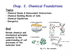
Chap. 2. Chemical Foundations. Topics Chemical Bonds & Noncovalent Interactions Chemical Building Blocks of Cells Chemical Equilibrium Energetics. Goal Review chemical and biochemical principles essential to the understanding of cell processes at the molecular level. Fig. 2.1. Key concepts.

E N D
Chap. 2. Chemical Foundations • Topics • Chemical Bonds & Noncovalent Interactions • Chemical Building Blocks of Cells • Chemical Equilibrium • Energetics Goal Review chemical and biochemical principles essential to the understanding of cell processes at the molecular level. Fig. 2.1. Key concepts
Review of Covalent Bonding The most abundant elements in cells are H>O>C>N>P>S. The number of covalent bonds formed by these elements is shown in Table 2.1. Note that oxygen and nitrogen have unshared pairs of electrons in bonding orbitals. The most common bonding orbitals for carbon (sp3, tetrahedral; sp2, trigonal planar) are shown in Fig. 2.3 (right). When 4 different substituents are bonded to sp3 carbon, the carbon is asymmetrical. These carbons are chiral and optically active.
Properties of Water Molecules Water is a polar solvent. It readily dissolves polar and ionic compounds, but not nonpolar hydrocarbons. Water molecules are polar because hydrogen and oxygen atoms have substantially different electronegativities (affinities for electrons) (Fig. 2.5). Because electrons are shared unequally, the -O-H bonds are dipolar and partial positive and negative charges occur on H and O. This feature plus the fact that 2 unshared pairs of electrons are located at the top of these sp3 hybridized molecules creates a net dipole moment. Water dipoles interact well with dissolved polar and ionic solutes.
Noncovalent Interactions (Bonding) Noncovalent interactions are weak electrical bonds between molecules. Types of noncovalent interactions are 1) ionic (electrostatic) bonds, 2) H-bonds, and 3) van der Waals interactions. Noncovalent interactions (1-5 kcal/mol) are typically ~100-fold weaker than covalent bonds (Fig. 2.6). Their stability is only slightly greater than thermal energy in biological systems. Nonetheless, noncovalent interactions play important roles in protein and nucleic acid stabilization because they are "collectively strong." Note that the hydrophobic effect drives molecular interactions, but is not a noncovalent bond per se.
Ionic Interactions Ionic interactions occur between cations and anions. These bonds are non-directional, and strength depends on the distance of separation (r) according to 1/r2. Strength also depends on the medium (dielectric constant), and is less in polar than nonpolar solvents. Ionic compounds such as NaCl are readily dissolved n water (Fig. 2.7). Solvation spheres of water molecules surround ions in solutions. Water molecules orient so that the negative ends of their dipoles contact cations and the positive ends contact anions in solution.
H-bonds H-bonds are noncovalent interactions occurring between the H atom of a dipolar molecule such as water, and the unshared electron pair of another atom (i.e., O or N). These bonds represent the primary way in which water molecules interact with themselves and many types of biomolecules (Fig. 2.8). H-bonds are highly directional in that strength depends on the proper alignment of the interacting atoms. Directionality confers bonding specificity as with the Watson-Crick H-bonds between the bases of double helical DNA.
van der Waals Interactions van der Waals interactions are bonds between fluctuating, induced dipoles within the electron clouds of interacting molecules. These bonds can occur between nonpolar or polar molecules. van der Waals bonds are extremely dependent on the distance of separation between molecules, and are significant only when the electron clouds of the molecules are just touching. van der Waals interactions are demonstrated for two O2 molecules in Fig. 2.10. The covalent and van der Waals radii are shown.
The Hydrophobic Effect The hydrophobic effect refers to the entropy-driven aggregation of nonpolar molecules in aqueous solution that occurs to minimize the ordering of water molecules with which they are in contact. This is not an attractive force, but rather a thermodynamically driven process. Fig. 2.11 shows the cage-like structures formed by water molecules surrounding a nonpolar solute. The hydrophobic effect drives the formation of membranes and contributes to the folding of proteins and the formation of double helical DNA.
Molecular Complementarity Structurally complementary molecules can bind to each other (Fig. 2.12). Binding often is mediated solely by noncovalent interactions. The extent of molecular complementarity sets binding affinity (how strongly molecules interact) and specificity (which molecules interact). Affinity and specificity are very important for molecular function. Intramolecular complementarity establishes the 3D shapes of proteins, etc.
Intro to Biological Macromolecules As shown in Fig. 2.13, biological macromolecules are polymers that are assembled from "building block" monomer units. In all cases, polymerization occurs via condensation reactions in which water is eliminated.
Eukaryotic Cell Membranes Eukaryotic cell membranes are composed of molecules such as phospholipids (Fig. 2.13), cholesterol, and proteins. Phospholipid bilayers are noncovalent assemblies of phospholipids. The hydrophobic fatty acyl chains of phospholipids aggregate together in bilayers. The two layers of a bilayer are called leaflets. Bilayers are mostly impermeable to polar and ionic molecules, but allow nonpolar compounds to pass through. Biomembranes typically contain other lipids such as sterols and glycolipids, and proteins.
Amino Acids There are 20 "standard" amino acids that are specified by the genetic code and polymerized into proteins by ribosomal translation. Amino acids contain an a-carbon, to which typically 4 different substituent groups are attached (Fig. 2.4). These groups are the a-amino group, the a-carboxyl group, hydrogen, and the variable R-group (side-chain). The a-amino and a-carboxyl groups are charged at neutral pH. There are two possible configurations for these groups--the "D" and "L" stereoisomers, which are mirror images of each other (enantiomers). The standard amino acids have the L-configuration. Amino acids are classified based on the characteristics of their R-groups (Fig. 2.14, next three slides).
The Hydrophobic Amino Acids With the exception of tyrosine, these amino acids lack polar functional groups within their side-chains. Hydrophobic amino acids often are found in the interior of proteins. The hydroxyl group of tyrosine residues exposed at the protein surface can be phosphorylated in receptors, etc.
The Hydrophilic Amino Acids These amino acids contain charged or H-bonding functional groups within their side-chains. For this reason, they often are found at the surface of proteins. The R-groups of lysine and arginine are positively charged at neutral pH. The R-groups of aspartate and glutamate are negatively charged. The histidine R-group may or may not be positively charged depending on its environment. Serine and threonine can be covalently modified by phosphorylation or glycosylation.
Other Amino Acids These amino acids carry out special functions in proteins. Glycine and proline often are found in "turns" (bends) within proteins. Cysteine can be modified with lipid groups or covalently bound to another cysteine in a "disulfide bond". Disulfide bonds (bottom right) contribute to protein folding stability. Disulfide bond
Properties of Nucleotides Nucleotides are the building blocks of nucleic acids (DNA & RNA). Nucleotides are composed of a base, a sugar, and one or more phosphates (Fig. 2.16a). A nucleoside contains just the base plus the sugar. The sugar is ribose in RNA and 2-deoxyribose in DNA (Fig. 2.16b). The most common bases are the purines, adenine and guanine, and the pyrimidines, uracil, thymine, and cytosine (Fig. 2.17). The naming of nucleosides and nucleotides is described in Table 2.3.
Properties of Carbohydrates The most common monosaccharides in cells contain 5 (pentoses) or 6 (hexoses) carbons. Monosaccharides are classified as aldoses or ketoses, depending on whether they contain an aldehyde or ketone group. Pentoses and hexoses typically form rings, as illustrated for the most common monosaccharide, glucose (Fig. 2.18a). Aldohexoses occur in 16 stereoisomeric forms. Dimers of monosaccharides are called disaccharides, and polymers are called polysaccharides. Common disaccharides are lactose (galactose-glucose) and sucrose (glucose-fructose). Common polysaccharides formed from glucose are glycogen in animals, and starches and cellulose in plants. The linkages between monosaccharide units are called glycosidic bonds.
Properties of Fatty Acids Fatty acids contain a polar carboxylic acid group and a hydrocarbon tail of variable length (14-20 carbons in animals) (Table 2.4). Fatty acids are further classified as saturated or unsaturated depending on their content of double bonds (usually cis in animals). cis double bonds introduce kinks into the tail (see Fig. 2.21), perturb packing interactions, and increase the fluidity of membrane lipids. The carboxyl group is joined to glycerol via ester (acyl) linkages in triacylglycerols and phosphoglycerides.
Phospholipids Phospholipids are the most prevalent components within biomembranes. In phosphoglycerides, two fatty acids are linked to glycerol at carbons-1 and -2. A phosphate and polar group (such as choline) are linked to carbon-3 (Fig. 2.20). The phosphate and polar group constitute the "head group.” The head groups of phospholipids commonly found in eukaryotic cells are shown in Table 2.5.
Equilibrium Constants A reaction, aA bB, is at equilibrium when the rate of the forward (vf = kf[A]a) and reverse (vr = kr[B]b) reactions are the same. The equilibrium constant, Keq, is defined as Keq = kf/kr = [B]b/[A]a Note that the steady-state concentrations and ratios of the components of a reaction occurring within a cell often are very different from the concentrations and ratios of the components when the reaction is at equilibrium (Fig. 2.23). C X
Dissociation Constants Many proteins bind to molecules called ligands. Ligands can be small molecules, proteins, or other biopolymers, such as DNA or polysaccharides (Fig. 2.24). Dissociation constants (Kds) are used to measure the affinity of protein-protein and protein-DNA interactions, for example. Kds are equivalent to the inverse of the Keq for a reaction, as illustrated in the following equation. Protein (P) + DNA (D) Protein-DNA (P.D) Kd = [P] [D] / [P.D]
Chap. 2 Meaning of the Kd Example: Protein (P) binding to DNA (D) P + D P.D Kd = [P][D]/[P.D] What is the ratio of [D]/[P.D] for different values of [P]? [P] = 0.1 x Kd [D]/[P.D] = 10/1 [P] = Kd [D]/[P.D] = 1/1 [P] = 10 x Kd [D]/[P.D] = 1/10 This shows that the DNA binding site is about 10% occupied when the concentration of [P] is 10-fold lower than the Kd, 50% occupied when [P] is the same as the Kd, and 90% occupied when [P] is 10-fold greater than the Kd.
Solution pH Water has a small but measurable tendency to ionize H2O H+ + OH- In pure water, [H+].[OH-] = 1 x 10-14 M2. Thus, in a neutral solution [H+] = [OH-] = 1 x 10⁻⁷ M. pH is defined as -log [H+]. Thus, pH = 7.0 for a neutral solution. Acidic solutions have pH < 7.0 and basic solutions have pH > 7.0. Remember, [H+] changes by 10-fold for a 1 unit change in pH. pH affects the properties of nearly all biomolecules. The pHs of a number of solutions is shown in Fig. 2.25.
Ionization of Weak Acids Biomolecules containing carboxylic acid groups are weak acids. Unlike strong acids (e.g., HCl), weak acids are << 100% dissociated in water. The dissociation of a weak acid is written as HA H+ + A- for which the acid dissociation constant is Ka = Keq = [H+] [A-] / [HA] Henderson-Hasselbach Equation Another form for the equilibrium equation is pH = pKa + log [A-]/[HA] Examination of this equation shows that [A-]/[HA] = 10/1 when pH = pKa + 1 [A-]/[HA] = 1/1 when pH = pKa [A-][HA] = 1/10 when pH = pKa - 1
Relationship Between pH, pKa, and Acid Dissociation The percentages of the conjugate acid (H2CO3) and conjugate base (HCO3-) forms of carbonic acid are plotted as a function of pH in Fig. 2.26. At pH extremes, one or the other species makes up essentially 100% of the solution. At the pKa, a 50/50 ratio of the two forms is present. The same type of information can be derived from a titration curve (next slide).
Titration of Acetic Acid In a titration, the conjugate acid form (HA) of a weak acid is stoichiometrically converted to its conjugate base form (A-) by the addition of a strong base. The titration curve for acetic acid (Fig. 2.27) can be used to explain the HH equation. The ratios of [A_]/[HA] are indicated as a function of pH. The optimum buffering range of acetic acid is +/- 1 pH unit from the pKa. [A-]/[HA] = 10/1 [A-]/[HA] = 1/1 [A-]/[HA] = 1/10
Behavior of H3PO4, a Triprotic Weak Acid H3PO4 has 3 dissociable protons, 3 pKas, and 3 plateaus on its titration curve (Fig. 2.28). The midpoint of each plateau occurs where the pH is equivalent to one of the pKas. The cell cytosol is buffered mostly by phosphate, namely the H2PO4-/HPO42- conjugate acid/conjugate base pair. The ratio of these species is 1/1 at pKa2. pKa2
Free Energy Changes for Reactions The free energy of a system or chemical compound is denoted by "G". The free energy change (∆G) for a reaction (Reactants Products) is calculated using the equation ∆G = GProducts - GReactants If ∆G < 0, the reaction will go to the right. If ∆G = 0, the reaction is at equilibrium, and goes neither direction in a net sense. If ∆G > 0, the reaction will go to the left. Note that the term ∆G0' is used for reactions occurring under standard biochemical conditions, where T = 298˚K, P = 1 atm, and the starting concentrations of all components (other than H+) is 1 M. For [H+], pH = 7.0. Units of G are kcal/mol.
∆Gs for Reactions (cont.) Gibbs free energy equation The ∆G for a reaction is determined by the enthalpy change (∆H) and entropy change (∆S) for the reaction. ∆G = ∆H - T ∆S ∆H reflects changes in the chemical bonds for all molecules in the reaction. ∆S reflects changes in the entropy (randomness) of all components participating in the reaction. If ∆G < 0, the reaction is exergonic, and will proceed spontaneously to the right. If ∆G > 0, the reaction is endergonic and will not proceed to the right without an investment of energy. ∆G is always negative when ∆H < 0 and ∆S > 0. Conceptual views of exergonic and endergonic reactions are shown in Fig. 2.29.
Calculation of ∆G Under Any Condition The calculation of ∆Gs for reactions under any concentration conditions (e.g., inside cells) is performed using the equation ∆G = ∆G0' + 2.303 RT log Q where Q = [products]/[reactants] (the mass action ratio) inside the cell, etc. In other words, ∆G is equivalent to the standard free energy change (∆G0') plus energy (positive or negative) resulting from differences in the concentrations of components from 1 M (standard conditions). Depending on the magnitude of Q, a reaction can proceed in the opposite direction from that under standard conditions. Note that R = 1.987 cal/˚K-mol.
Relationship Between ∆G0' and Keq When a reaction is at equilibrium, substitution of ∆G = 0 into the previous equation shows that ∆G0' = - 2.303 R T log Keq (Here Q = Keq = [products]/[reactants]). This equation is the same as Keq = 10 -∆G0'/2.303RT An inspection of the equations shows that ∆G0' < 0 when Keq > 1 ∆G0' = 0 when Keq = 1 ∆G0' > 0 when Keq < 1
Energy Coupling in Biological Reactions Endergonic reactions of biochemistry very often are driven forward by coupling them to the hydrolysis of ATP. As long as ∆G1 + ∆G2 = ∆Gsum < 0 the reaction will go forward. g b a Hydrolysis of either of the two phosphoanhydride bonds in ATP (Fig. 2.31) releases -7.3 kcal/mol of energy, which is more than enough energy to drive most endergonic reactions forward. Hydrolysis of the phosphoester linkage between the a-phosphate and ribose releases only -2.2 kcal/mol, and typically is not used for energy coupling purposes.
Mechanisms of Energy Coupling Assume that ∆G > 0 for reaction B + C D. Very often such a reaction can be driven forward via a high energy intermediate created via ATP phosphorylation. B + ATP B-p + ADP B-p + C D + p B + C + ATP D + ADP + p B-p serves as a common intermediate in the two reactions which go forward because ∆Gsum < 0. ATP (or GTP) hydrolysis also can provide energy for reactions via the creation of a high energy conformational state in a protein (e.g., myosin) carrying out an endergonic reaction.