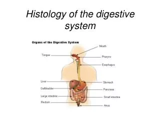
Download

1 / 23
280 likes | 600 Vues
Histology of the Skin. Kelli A. Hutchens, MD, FCAP Director of Dermatopathology Assistant Professor Loyola University Medical Center Assistant Director of Mechanisms of Human Disease Loyola Stritch School of Medicine. Objectives.

E N D
Histology of the Skin Kelli A. Hutchens, MD, FCAP Director of Dermatopathology Assistant Professor Loyola University Medical Center Assistant Director of Mechanisms of Human Disease Loyola Stritch School of Medicine
Objectives • Identify the epidermis, dermis, and subcutis of the skin • Name and label the layers five layers of the of the epidermis • Compare and contrast the anatomic and histologic differences between thick and thin skin • Identify (when possible) / or know the location of the following cells: • Keratinocyte • Melanoycte • Merkel cell • Langerhan cell • Describe the general function and location of the following components of the dermal epidermal junction and intercellular space. • Hemidesmosomes • Basement membrane • Basal layer keratinocytes • Anchoring fibrils • Desmosomes • Name and identify the two regions of the dermis • Identify and classify the following: • Eccrine gland • Sebaceous gland • Apocrine gland • Compare and contrast the histologic appearance, function, and location of Pacinian corpuscles versus Meissner’s corpuscles.
Overview of the Skin Epidermis Dermis Subcutis
Epidermis • Most superficial layer of the skin • Composed of multiple layers of keratin containing epithelial cells = keratinocytes with Melanocytes, merkel cells, and Langerhans dispersed throughout • Major functions: • Maintenance of fluid and electrolyte balance • Protection from ultraviolet light • Sensory and immune function
Epidermis : Layers • Cancel Lab Get Some Beer Stratum Corneum Stratum Lucidum Stratum Granulosum Stratum Spinosum Stratum Basale Thin Skin Thick Skin Rete ridge Stratum Lucindum
Differences between thin & thick skin Thin Skin Thick Skin Entire body except thick skin areas. Less than 5 layers of stratum corneum with no stratum lucidum Hair follicles present except lips, labia minora, and glans penis • Palms of hands and soles of feet = acral skin • 5 layers thick stratum corneum with increased granular layer • More sensory receptors • Lack sebaceous glands and increased eccrine glands • No hair follicles
Epidermis • Desquamatization: • Layers of epidermis represent vertical maturation from undifferentiated basal cells to fully differentiated cornified cells • From basal cell to cornified cell takes about 25 days • Shorter maturation periods seen in inflammatory conditions such as psoriasis • Keratin production also changes as the cell matures and disruption in the mechanism can effect the integrity of the keratinocytes such as in Haily-Haily and Darier’s Disease.
Epidermis Cell to Cell Adherence Zona occludens: tight junctions prevent diffusion across cells Zona adherens: Ca++ dependent cadherins that connect to actin Macula adherens: Made of desmosomes Gap junctions: communication for electric / metabolic function Hemidesmosomes: connect cells to BM Basement Membrane
Epidermis Desmosome = Intercellular Bridges
Epidermis: Melanocytes Melanocytes: clearish cells in basal layer with dark nuclei ; ratio of 1: 10. Langerhans’s Cells: dendritic cells of the epidermis. Sit in the mid-spinous. Not visible by light microscopy. Merkel Cells: located in the stratum basale. Also not visible by light microscopy. They are receptor cells that establish synaptic contacts with sensory nerves and contain granules of neurotransmitters.
Dermal-Epidermal Junction • Connects the epidermis and dermis • It is composed of proteins which provide a firm connection • Hemidesmosome: connects basal keratinocytes to basement membrane • Basement membrane • Lamina lucida = collagen types XVII, XIII, laminin 5 & 6 • Lamina densa = collagen type VII • Anchoring fibrils attach the basement membrane to the dermis hooking on to collagen VII and collagen I.
Basement Membrane Hemidesmosomes Basal layer keratinocytes of epidermis Laminins 5 & 6 Lamina Lucida Basement Membrane Lamina Densa Collagen Type VII Collagen type XVII, XIII Anchoring Fibrils Dermis Collagen type I
Dermis • Everything below the dermal epidermal junction / basement membrane • Connective tissue layer with contains blood vessels, nerves, sensory receptors, adnexal structures
Dermis • Two layers • Papillary dermis = includes the dermal papilla which project into the epidermis • The increases contact area preventing epidermal detachment • Also results in an undulating pattern which vary by anatomic location and individual resulting in grooves in the epidermis =dermatoglyphics (fingerprints) • Capillaries, free nerve endings and encapsulated sensory receptors called Meissner’s corpuscles. • Reticular dermis = area between the papillary dermis and subcutis
Papillary Dermis Capillaries Papillary Dermis
Dermis • The dermis is composed of two major types of fibers: • Type I Collagen • Elastic fibers: three types based on microfiber and elastin content
Sebaceous Glands Reticular Dermis Erector Pili muscle Hair Follicle
Dermal Appendages Sebaceous Glands Hair Follicle Eccrine Glands Pilar Muscle
Sebaceous Glands • Usually associated with hair follicles • Simple branched acinar glands • Several acini that empty into single duct • Holocrine secretion • Empty “sebum” into hair follicle
Hair Follicle cross section (above the level of the bulb) Connective Tissue Sheath Outer Root Sheath Inner Root Sheath Hair Cuticle Hair Cortex Bulb Hair Medulla Papilla Matrix
Eccrine Glands • Merocrine sweat glands • Release to adjust body temperature • Three cell types • Dark cells: pyramid shaped with secretory granules line lumen of tubule • Clear cells: located toward basement membrane • Myoepithelial cells: spindle shaped contractile cells
Apocrine Glands • Apocrine glands • Similar to eccrine glands but larger lumens and ducts empty onto superficial regions of hair follicle • Release product by shedding of part of cytoplasm = apocrinesnouting • Influenced by hormones (sexual scent glands) • Only found on axilla, areola, perianal and genital area
Subcutis • Subcutis • Area deep to the dermis • Includes the hypodermis • Loose connective tissue containing adipose tissue, nerves, sensory receptors, arteries and veins • Provides a flexible attachment to the underlying muscle and fascia Pacinian Corpuscle Adipocytes Hair bulb in the subcutis of the scalp.