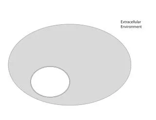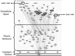
The Extracellular Space
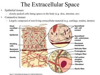
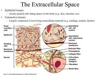
The Extracellular Space. Epithelial tissues closely packed cells lining spaces in the body (e.g. skin, intestine, etc) Connective tissues Largely composed of non-living extracellular material (e.g. cartilage, tendon, dermis). The Extracellular Space.

The Extracellular Space
E N D
Presentation Transcript
The Extracellular Space • Epithelial tissues • closely packed cells lining spaces in the body (e.g. skin, intestine, etc) • Connective tissues • Largely composed of non-living extracellular material (e.g. cartilage, tendon, dermis)
The Extracellular Space • Proteins in plasma membrane have sugars attached • Glycocalyx • Mediate cell interactions • Provide mechanical support • Barrier to particles • Binding sites for regulatory factors
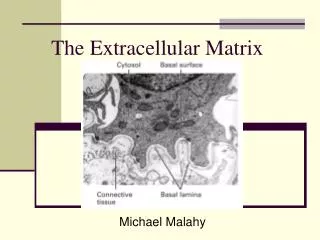
The Extracellular Matrix (ECM) • An organized network of materials located beyond the plasma membrane
The Extracellular Matrix (ECM) • Basement membranes • Thick regions of ECM • Surround muscle/fat • Underlie the basal surface of epithelial tissues
The Extracellular Matrix (ECM) • Basement membranes • Separate different tissues • Provide mechanical support • Barrier to macromolecule and cellular movement • Substrate for cell migration • Generate signals that maintain cell survival
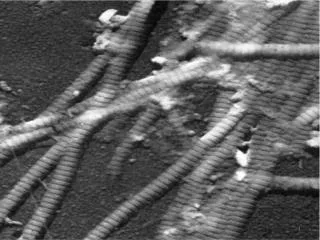
The Extracellular Matrix (ECM) • Collagens (27 different types) • High tensile strength (resistant to pulling forces) • Alpha-helical trimers bundle together into fibrils • Types I, II, III (fibrillar) form rigid cables • Adjacent collagens are strengthened by covalent cross-links • Hydroxylysine - lysine • Type IV (nonfibrillar) can form an interconnected lattice
The Extracellular Matrix (ECM) • Collagens (27 different types) • Type IV (nonfibrillar) can form an interconnected lattice • Composed of helical and non-helical segments (flexibility) • Globular domains at each end (lattice contact points) • Collagens bind: • Fibronectins • Integrins (cell surface)
The Extracellular Matrix (ECM) • Diseases caused by defects in collagen genes • Osteogenesis imperfecta • Fragile bones • Ehlers-Danlos syndrome • Hyperflexible joints, highly extensible skin
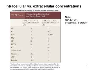
The Extracellular Matrix (ECM) • Proteoglycans • Protein core + glycosaminoglycan (GAG) polysaccharide complex • Chondroitin sulfate & keratin sulfate • High amount of negative charge binds cations and H2O • Hydrated gel resists compressive forces • Hyaluronic acid links many proteoglycans to form extremely large molecules • Fill the scaffold created by collagens
The Extracellular Matrix (ECM) • Fibronectins • Modular domains for interactions • Bind collagens, proteoglycans, integrins at cell surface • Important for: • linking ECM components together, cell attachment to matrix, cell migration NC cells
The Extracellular Matrix (ECM) • Laminins • 3 polypeptides linked by disulfide bonds • Form a second lattice interwoven with Collagen IV lattice • Bind to proteoglycans, integrins at cell surface PGC on laminin
ECM Remodeling • Matrix metalloproteinases (MMPs) • Enzymes that degrade ECM proteins • Tissue remodeling • Cell migration • Wound healing
Steps leading to metastatic spread MMP activity
Cell - ECM Interactions • Integrins • Only found in animals • Heterodimer of alpha and beta subunits • 18 alpha and 8 beta subunits known • 12 different alpha/beta combinations known • Transmembrane proteins • Extracellular domain, transmembrane domain, intracellular domain • Inside-out signaling • Post-translational alterations to cytoplasmic tail regulate conformation changes in extracellular domain • Talin separates beta from alpha to open receptor to active state Plasma membrane talin
Cell - ECM Interactions • Ligand binding • RGD loop of Fibronectin binds to integrin receptor extracellular domain • Isolated RGD Loop can be exploited to block platelet aggregation / blood clotting
Cell - ECM Interactions • Integrins • Two major functions • Adhesion to substrate • Receptors cluster increasing overall strength • Signal transmission • Binding of ligand (collagen) can change cytoplasmic domain • Cytoplasmic domain can activate kinases such as FAK and Src • Activated kinases can transmit signals to nucleus and change gene expression
Cell - ECM Interactions • Structures important for adhesion to substrate • Focal adhesions: • Scattered, discrete, transient, dynamic, rapidly form and break • Clusters of integrins bound to collagen / Fibronectin • Cytoplasmic domains attach to cytoskeleton connecting exterior forces to internal signals • Actin filaments • Focal adhesion kinase (FAK)
Cell - ECM Interactions • Structures important for adhesion to substrate • Hemidesmosome • more permanent anchor to basement membrane • Integrins bound to laminin to dense collection of intermediate filaments
Cell - ECM Interactions • Structures important for adhesion to substrate • Hemidesmosome • Disease: epidermolysis bullosa • Epidermis poorly connected to basement membrane / dermis • Fluid accumulates in between = blister (keratins)
Cell - Cell Interactions • Cadherins: Ca2+ dependent adhesion • Homophilic interactions allow self-sorting of mixed cell populations • Disease role: metastasis of cancer • Lose adhesion by downregulating cadherin expression • Penetrate / invade barriers by upregulating MMP expression
Cell - Cell Interactions • Structures important for cell-cell adhesion • Adherens junctions (30nm gap between cells) • Cadherin-cadherin interactions in belt-like strips holding two cells together • Cytoplasmic domains link via beta-catenin and alpha-catenin to the cytoskeleton
Cell - Cell Interactions • Structures important for cell-cell adhesion • Desmosomes (1 um diameter disc) • Resist mechanical stress • Cadherin-cadherin interactions linked to cytoskeleton (intermediate filaments)
Cell - Cell Interactions • Tight junctions • Seal two membranes together • Block paracellular movement • Occludin and claudins (24 genes) • Different claudins have different permeabilities • #1 doesn’t allow H2O to pass, #16 is permeable to Mg2+ • Important for maintaining blood-brain barrier
Cell - Cell Interactions • Gap junctions • Join cytoplasmic spaces between adjacent cells via a narrow pore • 1.5nm diameter • 1kD cutoff, small molecules freely pass (ATP, cAMP, Ca2+, etc) • Subunits are connexins • Open / close regulated by phosphorylation • Integrates cells of a tissue into a functional unit
Plant cell-cell interactions • Plasmodesmata • Join adjacent plant cytoplasmic spaces • Capable of dilation, 1kD cutoff can open to a 50kD cutoff • Exploited by some plant viruses
Roles of the plant cell wall • Cell wall functions • Structural role supporting and protecting plant cells • Cellulose microfibrils confer tensile strength • Signaling roles • Cell wall-associated transmembrane protein kinases • Dynamic not static, undergoes significant remodeling