Download
1 / 92
1.11k likes | 4.66k Vues
Complications of Cardiac Catheterization. Grossman’s cardiac catheterization, angiography, and intervention Chapter 3 CV R4 陳柏升 Supervisor: Prof. 蔡良敏 . Introduction. The risk of producing a major complication ― less than 1% during most procedure type currently

E N D
Complications of Cardiac Catheterization Grossman’s cardiac catheterization, angiography, and intervention Chapter 3 CV R4 陳柏升 Supervisor: Prof. 蔡良敏
Introduction • The risk of producing a major complication ― less than 1% during most procedure type currently • The risks of complication for the individual patient • Demographics (age, gender) • The cardiac anatomy (left main CAD, AS, LV dysfunction) • The clinical situation (ACS, shock, ARF) • The type of procedure being performed (diagnosis, PCI)
Introduction • Familiarity with those risks can be of immeasurable value in the following: • Anticipating increased risk of complication • Taking extra precautions to avoid them (TPM for PCI) • Promptly recognizing complications when they occur • Taking corrective and potentially life-saving action
Introduction • Discussed with the patient and family • The detailed of the planned procedure and its anticipated risks • Informed consent • Documented in the patient’s chart • Specify the type of procedure that is planed • The potential major complications and their estimated risk of occurrence
Introduction • Should be intimately knowledgeable about the potential complications of the procedures • Collect information about the frequency of these complications on at least a yearly basis and review those data with physician staff
Death Diagnostic catheterization • Incidence • The first Society for Cardiac Angiography registry from 1979 to 1981: 0.14% procedure-related mortality • The second registry from 1984 to 1987: slightly more to 0.1% • The third registry in 1990s: 0.08%, with a 1.5% incidence of any major complications • High risk groups
Death Diagnostic catheterization • Severe left main disease • More than 20 times higher than patients with 1-V-D • Careful engaging and “puff” test in RAO and caudal angulation view to screen for left main disease • If ostial left main stenosis is present, AP injection may be performed → RAO and cranial angulation to see LAD and diagonal branches • More view offers little information and increase the risk fo triggering the vicious cycle to irreversible collapse • IABP for unstable patient and arrange for bypass surgery
Death Diagnostic catheterization • Severe left ventricular dysfunction (EF < 30%) • Seven-fold increased risk • Particularly is associated with PCWP > 25mmHg and systolic BP < 100 mmHg • An effort to make CHF under control before cath • Right heart catheterization should always be performed to get more information • PWCP > 30mmHg, every effort made to improve hemodynamic status • MAP > 65 mmHg → diuretics, oxygen supply, vasodilator • MAP < 65 mmHg → a positive inotrpic agent • IABP • Percutaneous cardiopulmonary bypass (CPS) • low-osmolar-contrast agent to produce less myocardial depression
Death Diagnostic catheterization • Patients who have previously undergone CABG • Older, more diffuse coronary and generalized atherosclerosis, worse left ventricular function, more length and complex procedure • The Post CABG trial: 2635 diagnostic angiogram; no death, 0.7% major complications • Pediatric patients ― high risk group • Potential explanations for declining mortality rate • Improvement in catheter design, imaging systems, contrast agents • High annual proceduce volume ( >1.5 million/year) • Shorter procedure time • Heparin use?
Death Intervention procedures • More higher mortality in intervention because • More aggressive catheters • Superselective cannulation of diseased coronary arteries • Brief interruption of coronary and systemic flow • Roughly 10-fold higher than purely diagnostic procedure (1% : 0.1%) • A wide variation in risk based on patient comorbidities, clinical condition, and procedure type
Myocardial infarction ― Diagnostic catheterization • Transient myocardial ischemia is relatively common during diagnostic catheterization and coronary intervention • Respond promptly to drug therapy • Deflation of the angioplasty balloon
Myocardial infarction ― Diagnostic catheterization • Myocardial infarction is an uncommon but important complication of diagnostic cardiac catheterization • Reduction progressively • 0.25% in the late 1970s to 0.05% currently • Causes of reduction • Experience ― greater attention to catheter flushing, pressure damping • Heparin use for angioplasty • High risk groups ― patient-related and technique-related • The extent of vessel diseases: 1-V-D : 3-V-D : left main → 0.06% : 0.08% : 0.17% • The clinical indication (unstable angina or recent subendocardial infarction) • IDDM • “Angioplsty stand-by” for these unstable patients
Myocardial infarction ― Diagnostic catheterization • IABP or remaining intravenous heparinization for patients whose severe lesions are suitable for bypass surgery • Unstable patient should not reversed heparinization with prostamine, absent a life-threatening bleeding complications • Resume heparinization in 2 hours later after sheath is removed • Post-procedure MI is rare in patients who complete their catheterization without unstable signs • Should severe ischemic instability develop after the patient leaves the cath lab, aggressive therapy in indicated → coronary intervention or bypass surgery
Myocardial infarction ― Interventional procedure • Mechanisms • Dissection • abrupt vessel closure • “snow-plow” occlusion of side branches • Spasm or no-reflow • Thrombosis • distal embolization • Incidence rate decreased progressive, the current experience suggested that emergent bypass surgery and Q-wave infarction rates is 1% • Some elevation of CPK and CK-MB ― at least 20% of patients with otherwise successful intervention • Low order (1 to 3 times the upper limit of normal) appears to have no short- or long-term consequences • Only elevation 5 times above normal tends to adversely impact late survival ― views as a major complication, equivalent to Q-wave infarction
Cerebrovascular complications • Uncommon but potentially devastating complications • Decrasing incidence: 0.23% (1973) → 0.07% (current) • embolic in origin • Most cases • Many emboli are dislodged from unsuspected aortic plaque or diffuse atherosclerosis; emboli originate from cardiac chambers, thrombotic coronary arteries, or surface of cardiac valves • Technical errors: sloppy catheter flushing, introducing of air bubbles during contrast injection, inadvertent placement of wires and catheters into arch vessels, prolonged (> 3mins) wire dwell time, etc. • Avoid • “catching” in aortic surface • Dislodgeable mural thrombus • Avoid transseptal catheterization or mitral valvuloplasty in patients with left atrial thrombus, which may increase the incidence of stroke
Cerebrovascular complications • embolic in origin (cont.) • In patients with right-to-left shunt, paradoxical embolization may lead to stroke • Active left side endocarditis (AV or MV) : left side cath does not increase the incidence of embolic events (0/35)AJC 1979;44:1306 • Intracerebral hemorrhage • Patients receiving • Aggressive anticoagulation • Antiplatelet • Thrombolytic therapy • Neurologic consultation, image study (CT, MRI) • The distinction between embolic and hemorrhagic stroke is critical • Transient neurologic deficits • High-osmolar-contrast agents into the carotid and vertebral vessels
Local vascular complications • One of the most common problems • Problems including • Vessel thrombosis • Distal embolization • Dissection • Poorly controlled bleeding at the punctual site • poorly placed puncture • vessel laceration • excessive anticoagulation • poor technique in either suture closure or groin compression • Hemorrhage and hematoma ― evident within 12 hours; false lumen ― evident for days or even several weeks later
Local vascular complicationsDiagnostic catheterization • The Society for Cardiac Angiography registries ― 0.5 to 0.6% in incidence • Brachial approach • Arterial thrombosis • Causes • Formation of a thrombus in the proximal arterial part and failure to remove prior to repair • Secondary to an intimal flap within the arterial lumen • Secondary to local spasm • Preventions • Meticulous attention to the details of arterial repair • Adequate heparinization: systemic and local • Treatment • Fogarty catheter thrombectomy • Percutaneous transluminal angioplasty
Local vascular complicationsDiagnostic catheterization • Brachial approach (cont.) • Other complications • Injury to median nerve • Cutdown or compression by hematoma • Mild case: numbness and weakness for 3 to 4 weeks and return to normal: occasionally up to 6 months • Delayed dehiscence of arterial sutures with late arterial bleeding • Bacterial arteritis • Local cellulitis-phlebitis • Extensive soft tissue is dissected • Large vein are used and tied off • The catheterization procedure is long • Seroma and hematoma forms • Nonviable tissue is left in the incision • Poor surgical technique or violation of sterile procedure occurs
Local vascular complicationsDiagnostic catheterization • Femoral approach • Thrombosis (femoral artery) • Extremely rare, except a small femoral artery lumen (PAOD, DM, female), a large-diameter catheter or sheath (IABP) or long duration of catheter • S/S: leg pain or numbness, diminished distal pulse • Obstructive limb ischemia generally resolves and distal pulse returned when the sheath is removed • Ongoing complaint and diminished or absent distal pulsewith catheter removal → flow-obstructing dissection or thrombus → urgent vascular surgery →within 2 to 6 hours!! • Results in extension of thrombosis into smaller distal branch and muscle necrosis if delayed • Femoral venous thrombosis or pulmonary embolism • Rare (multiple venous lines or compression by large arterial hematoma etc.) but may be underreported: up to 10% asymptomatic positive lung perfusion scan • Continuous drip of heparinized saline t venous sidearm throughout the procedure to avoid this problem
Local vascular complicationsDiagnostic catheterization • Femoral approach (cont.) • Poorly controlled bleeding ― more common • Suggest laceration of the femoral artery • Try next-larger-diameter sheath or compressed manually until the procedure is completed • Reverse heparin and control bleeding with prolonged cpompression • Blood transfusion • Hematoma formation • usually resolve over 1 to 2 weeks • S/S: femoral nerve compression → quadriceps, weakness → takes weeks even months to resolve; surgical repair is not required generally • Hematoma may extend to retroperitoneal bleeding if puncture site is above inguinal ligament • unexplained hypotension, decreased Hct, ipsilateral flank pain; response to fluid challenge • best prevention
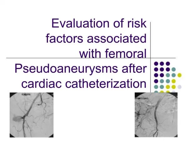
Local vascular complicationsDiagnostic catheterization • Femoral approach (cont.) • Pseudoaneurysm • Hematoma continuity with the arterial lumen • Blood flow in and out of the arterial puncture, expanding the cavity • pulsation, audible bruit, Duplex scan • Therapy • Surgical repair • transducer compress the neck for 30 to 60 minutes • procoagulant solutions or embolization coils with echo guiding • Prevention: accurate puncture of the common femoral artery and effective initial control of bleeding • A-V fistula • Not be clinically evident for days after procedure • Ongoing bleeding may decompress into the adjacent venous puncture site • To and fro continuous bruit • Surgical repair if fistula tends to enlarge with time or does not close within 2-4 weeks • High risks: low puncture site (superficial or profunda femoral arteries)
Local vascular complicationsInterventional procedure • A significantly high incidence of local vascular complications than pure diagnostic procedure ― 1 to 2% • Use of larger sheath • The intensity and duration of anticoagulation • Removal of the sheaths only after an overnight dwell • Various approaches for collagen plugging or percutaneous suture-mediated closure have been used • Avoid the discomfort of prolonged manual or mechanical compression • Allow early even immediate ambulation • Failed to demonstrate significant reduction of major vascular complications compared with compression
Arrhythmia or conduction disturbance • A variety of cardiac arrhythmia or conduction disturbance may occur ― VPCs, VT, VF, Af • Monitor the surface ECG with physiologic monitor with the pressure tracing and alarm system • The tools (defibrillator, pacemakers, ventilation support) and drugs to treat the rhythm disorder • All cardiac catheterization personnel must be fully certified in ACLS
Ventricular fibrillation • VPC and brief run VT(3-5 beats) are common • During the passage of catheter into the right or left ventricle • The offending catheter must be repositioned immediately • Other causes inducing ventricular fibrillation • Catheter transmission of “leakage” electrical current to the heart • Eliminated by the adoption of standards for grounding system, less than 20 μA between any two exposed conduction source • Intracoronary contrast injection • Most commonly with injection of ionic (high-osmolar) contrast agent into the right coronary artery, especially the injection is prolonged or the catheter pressure is damped • Change in injection technique and formulation of contrast agents reduce the incidence, 1.28%(1974) → <0.4%(1991)
Ventricular fibrillation • Other causes inducing ventricular fibrillation (cont.) • Patients with baseline prolongation of the QT interval • Some refractory ventricular ectopy is the setting of profound transmural ischemia or early myocardial infarction • Therapy • iv lidocaine (1.5 mg/kg over 1 minute, with a second bolus of 0.75 mg/kg 7 minutes later) • procainamide (15 mg/kg over 20 minutes,↓BP ,↑QRS or QT) • iv amiodarone (5 mg/kg over 20 minutes, 1 gm/24 hours)
Atrial arrhythmia • Atrial extrasystoles are common during • Catheter advancement from the right atrium to the SVC • Looping of the catheter in the right atrium to facilitate passage in a patient with enlargement of the right-side heart chamber • Usually subside once the catheter is repositioned • But may go on to Af or AF in sensitive patients • tends to revert spontaneously over a period of minutes to hours • Require additional therapy if they produce ischemia or hemodynamic instability
Atrial arrhythmia • Therapy • DC shock in both Af and AF with hemodynamic instability • Atrial flutter ― a brief (15 seconds) but rapid (300 -400 bpm) right atrial pacing • Ensue a stable atrial pacing location or triggering a VF since catheter migration into the ventricle • Atrial fibrillation ― may cause S/S with RVR, hypotension in patient with MS, hypertrophic cardiomyopathy, or diastolic left ventricular dysfunction • Rate control • iv beta blockers (inderal : 1mg ; esmolol : loading 500μg/kg/min for 30 sec, followed by 50~250 μg/kg/min) • iv CCB (verapamil 5mg) • Rhythm conversion • iv procainamide (15 mg/kg for 20 min) • ibutilide (on other QT-prolonging drugs; ↓K or Mg; bradycardia; QTc > 440 ms)> 60 kg : 1mg over 10 min ; within 4 hours, no other class III agents!!
Bradycardia • Occurs commonly during coronary angiography • At the end of a right coronary artery injection with ionic (high-osmolar) contrast agent • Forceful coughing • clear contrast from the coronaries • support aortic pressure and cerebral perfusion during asystole • restore normal cardiac rhythm • Vasovagal reaction • One of the most common complication (3% incidence) • bradycardia with hypotension, nausea, yawning, and sweating • triggered by pain and anxiety especially in hypovolemic status • 80% occurs as vascular access and 16% occur during sheath removal • adequate preprocedure sedation and local anesthetic
Bradycardia • Vasovagal reaction (cont.) • Treatment • Cessation of the painful stimulus • Rapid volume administration • Atropine • Additional pressor support may require if hypotension persist • May imply cardiac perforation when the pericardium is irritated by blood during catheter manipulation • Conduction disturbance • Uncommon but potentially serious cause of bradycardia during cardiac catheterization • Require no treatment except in the patient with preexisting bundle branch block ― asystole or cardiovascular collapse except escape rhythm takes over
Bradycardia • Conduction disturbance (cont.) • Complete AV block • Occurs during • Rotational atherectomy, especially in RCA or LCX • Aortic valvuloplasty • Treatment • Atropine is rarely helpful but should be given anyway since it has few adverse effects • Coughing • Pacing marker insertion • Prophylactic right-sided pacing catheters • Rotational atherectomy, especially in RCA or LCX • Aortic valvuloplasty
Perforation of the heart and great vessel • Rare • Heart perforation • 0.8% in 1968, RA-RV-LA-LV; 0.006% of diagnostic cath and 0.08% of coronary angioplasties now • Stiffer catheter and elderly women (>65 y/o) • S/S • Cardiac silhouette may enlarge • Pulsation of the heart borders on fluoroscopy may become blunted • Bradycardia and hypotension due to vagal stimulation • Cardiac tamponade ― elevation of RA pressure with loss with y descent • Paracentasis via subxyphoid approach and protamine infusion • Emergent surgical intervention? ― most will seal without surgery
Perforation of the heart and great vessel • Vessels perforation • Aorta: rare, except in the case of weakening by ascending aortic dissection or aneurysm • Pulmonary arteries • Rare; too stiff-tip guidewire or balloon inflated in a distal branch • Typically develop hemoptysis → require tamponade of the proximal pulmonary artery, embolization of the bleeding branch, double-lumen endotracheal tube, emergent lobectomy or pnemonectomy • Coronary artery • Unheared of diagnostic cath; the incidence rises to 1% with the advent of more aggressive new techniques for coronary intervention • Most are limited to deep injury to vessel wall; free perforation may leads to frank tamponade within seconds to minutes → seal the site of leakage by inflation of a balloon catheter, pericardiocentasis and protamine use if necessary, coil embolization, covered stent, emergent surgical intervention
Infection and pyogen reaction • Endocarditis prophylaxis and routinely antibiotics use are not recommended, except • performing a delayed intervention by exchanging sheaths that were placed in a earlier diagnostic procedure • Any break in sterile technique is suspected • Repeat procedure within 2 weeks → use contra-lateral groin • Increasing infection rate from the same groin • Full sterile precautions before procedure • Procedure from brachial approach • From femoral approach when the procedure is prolonged • The sheath was remain in place for any period • A stent and permanent pacemaker is being implanted • A vascular grafy is punctured • Guidelines of the Occupational Safety and Health Administration (OSHA): Sterile precautions for any procedure
Infection and pyogen reaction • Laboratory personnel • Vaccination for hepatitis B • Prophylactic zidovudine (AZT) for personnel being stuck by needle tainted with blood from HIV-positive patient • Avoid muitiuse drug vials and clean the room thoroughly between procedures • Post-procedure fever • Phlebitis • May develop after brachial catheterization • Low grade fever and a warm tender cord overlying the affected vein • Pyogen reaction • Shaking chills during or within the first hour • Fever spike as high as 102℉ • Caused by the presence of contaminating material that remain on incompletely cleaned catheter surfaces • Morphine use for symptom relief
Allergic and anaphylactoid reaction • Materials may cause allergic or anaphylactoid reactions • Local anesthesia • Iodinated contrast agent • Protamine • Preservatives • Skin testing with the intended agent at 1:1000 dilution if desired • Iodinated contrast agent • Most common in triggering allergic reactions ― up to 1% • Anaphylactoid reaction ― involve degranulation of circulating basophils and tissue mast cells by direct complement activation • Other clinical manifestations caused by histamine or other agents ― sneezing, urticaria, angioedema, bronchospasmwarm shock
Allergic and anaphylactoid reaction • Iodinated contrast agent (cont.) • High risk groups • Other atopic disorders • Allergy to penicillin • Allergy to seafood (contained organic iodine) • Prior reaction to contrast (as high as 15% to 35%) • Premedications ― reduce the incidence od a secone reaction to 5% to 10% and that of severe reaction to below 1% • Prednisolone ― 20mg tid for 24 to 48 hours • H1 antihistamine ― diphenhydramine 25mg tid • H2 blocker ― cimetidine or ranitadine • Nonionic contrast agent ― adds a further margin of safety • Record aortic pressure first when a patient have a history of well-documented prior severe contrast reactions • Emergent treatment ― intravenous injection of dilute epinephrine, 1:10,000 epinephrine • 1mL/minute until arterial pressure is restored • Avoid excessive doses ― may precipitate life-threatening hypertension, tachycardia or even VF
Allergic and anaphylactoid reaction • Prostamine • High risk groups • IDDM patients with NPH control (which contained with protamine) • Emergent treatment ― as previous described • Heparin-induced thrombocytopenia (HIT) ― rare • Definition • a fall in platelet count by at least 50% • Accomplished by a positive serologic test for the responsible antibody (usually IgG) • Platelet Fc receptor + PF-4/antibody → platelet activation • Onset is typically 7 to 10 days; bovine-derived more common than porcine-derived • Alternative anticoagulant • LMWH ― frequently cross-react with heparin antibody • Heparinoid (Organan, danaproid) • Direct antithrmbin compounds (hirudin, hirulog, or argatroban)
Renal dysfunction • At least 5% of patients experience a transient rise in serum creatinine greater than 1mg/dL following cardiac angiography • Precise mechanism of contrast-induced renal dysfunction ― not been established • High risk groups ― up to 50% • DM • Multiple myeloma • Volume depletion • Preexisting renal dysfunction • Patients who are receiving certain drugs(eg. GM, NSAID, ACE-I) • Most are non-oliguria, peak within 1 to 2 days and return to baseline by 7 days
Renal dysfunction • Fewer than 1% of patients go on to require chronic dialysis • Prospective trials comparing high and low-osmolar contrast agents have failed to showed consistent benefit • Main defense against contrast-induced nephropathy ― Limitation of total contrast volume to 3mL/kg • Adequate prehydration for any patients with impaired baseline renal dysfunction ― 12 hours before and after the contrast procedure
Renal dysfunction • Systemic cholesterol embolization • Another cause of renal failure following cardiac catheterization ― 0.15% of cathetherizations • High risk groups • Those with diffuse atherosclerosis • In whom insertion of a guiding catheter will frequently produce a shower of glistering particles on the table drape • Hallmarks of diagnosis ― evidence of peripheral embolization • Episodic hypertension or systemic eosinophilia may be apparent well before the other manifestations develops • Develops slowly ― over weeks to months • Half of the patients progressed to frank renal failure • Treatment ― purely supportive
Hypotension • One of the most common problem • Causes • Hypovolemia: inadequate hydration, blood loss, excessive contrast-induced diuresis • Reduction in cardiac output: ischemia, tamponade, arrhythmia, valvular regurgitation • Inappropriate systemic arteriolar vasodilation: vasovagal, excessive nitrate administration, response to contrast or inotrope-vasodilator drug • Right side catheterization for diagnosis and treatment • Initial empirical treatment and definite correction of hypotension and its causes before hypotension leads to secondary ischemia and irreversible spiral of left ventricular dysfunction
Hypotension • Treatment • Low filling pressure • Rapid volume administration • Atropine use if combined with inappropriate bradycardia • Look for potential site of blood loss • High filling pressure ― suggest primary cardiac dysfunction: ischemia, tamponade, VHD • Supported empirically by inotropic agents, vasopressor or circulatory devices • Immediate intervention or surgical intervention • Artificial pacing if bradycardia is present and is not response to atropine • Adequate airway maintenance • Coexistent sepsis, contrast reaction or and idiosyncratic vasodilator reaction to dopamine infusion
Volume Overload • Prone to fluid overload due to • Hypertonic contrast agents • Myocardial depression • Ischemia induced by contrast • Poor baseline left ventricular function • Supine position • Risk of contrast-induced renal dysfunction • Treatment • Optimizing volume status before or early in the procedure • Use of low-osmolar contrast agents • Supportive measures: inotropes, diuretics, vasodilators, IABP • Even more aggressive treatment is warranted once pulmonary edema develops, eg. intubation
Anxiety/Pain • Oral sedatives pretreatment(diazepam 5 to 10mg, diphenhydramine 25 to 50mg) and liberal use of local anesthesia at the insertion site • To understand why the patient is having pain first and whether anything to do to reverse the problem • The catecholamine surge associated with pain and anxiety may worsen the condition of a patient • Small dose of morphine, fentanyl, midazolam • Make the procedure more tolerable for both the patient and the staff • Monitor blood pressure, respiratory rate, and pulse oximetry • Antagonist drugs should also be stocked
Respiratory insufficiency • Not uncommon • Pulmonary edema • Baseline lung disease • Allergic reaction • Over sedation • Baseline ABG after sheath insertion • Low-flow supplemental oxygenation(2L/min) helps avoid episodes of desaturation • If oxygen consumption is to be measured as part of a calculation of cardiac output by the Fick method, oxygen administration should not be begun after that measurement
Retained equipment • Devices knot, become entrapped, or leave fragment in the circulation ― most of these events are precipitated when such devices are stressed beyond their design parameters • Operators should be familiar with device performance limits and be familiar with devices and techniques that can be used to recover the errant fragments, such as vascular snares, bioptomes, baskets, et.