The Urinary System
250 likes | 488 Vues
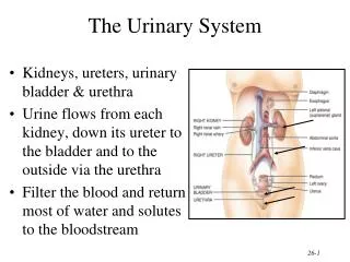
The Urinary System. Kidneys, ureters, urinary bladder & urethra Urine flows from each kidney, down its ureter to the bladder and to the outside via the urethra Filter the blood and return most of water and solutes to the bloodstream. Overview of Kidney Functions.

The Urinary System
E N D
Presentation Transcript
The Urinary System • Kidneys, ureters, urinary bladder & urethra • Urine flows from each kidney, down its ureter to the bladder and to the outside via the urethra • Filter the blood and return most of water and solutes to the bloodstream
Overview of Kidney Functions • Regulation of blood ionic composition • Na+, K+, Ca+2, Cl- and phosphate ions • Regulation of blood pH, osmolarity & glucose • Regulation of blood volume • conserving or eliminating water • Regulation of blood pressure • Release of erythropoietin & calcitriol • Excretion of wastes & foreign substances
Internal Anatomy of the Kidneys • Parenchyma of kidney • renal cortex = superficial layer of kidney • renal medulla • inner portion consisting of 8-18 cone-shaped renal pyramids separated by renal columns • renal papilla point toward center of kidney • Drainage system fills renal sinus cavity • cuplike structure (minor calyces) collect urine from the papillary ducts of the papilla • minor & major calyces empty into the renal pelvis which empties into the ureter
Blood & Nerve Supply of Kidney • Abundantly supplied with blood vessels • receive 25% of resting cardiac output via renal arteries • Functions of different capillary beds • glomerular capillaries where filtration of blood occurs • peritubular capillaries that carry away reabsorbed substances from filtrate (renal cortex) • vasa recta supplies nutrients to medulla • Sympathetic vasomotor nerves regulate blood flow by altering arterioles
Blood Vessels around the Nephron • Glomerular capillaries are formed between the afferent & efferent arterioles • Efferent arterioles give rise to the peritubular capillaries and vasa recta
The Nephron • Kidney has over 1 million nephrons composed of a corpuscle and tubule • Renal corpuscle = site of plasma filtration • glomerulus is capillaries where filtration occurs • glomerular (Bowman’s) capsule is double-walled epithelial cup that collects filtrate • Renal tubule • proximal convoluted tubule • loop of Henle dips down into medulla • distal convoluted tubule • Collecting ducts and papillary ducts drain urine to the renal pelvis and ureter
Cortical Nephron • 80-85% of nephrons are cortical nephrons • Renal corpuscles are in outer cortex and loops of Henle lie mainly in cortex
Juxtamedullary Nephron • 15-20% of nephrons are juxtamedullary nephrons • Renal corpuscles close to medulla and long loops of Henle extend into deepest medulla enabling excretion of dilute or concentrated urine
Structure of Renal Corpuscle • Bowman’s capsule surrounds capsular space • podocytes cover capillaries to form visceral layer • simple squamous cells form parietal layer of capsule • Glomerular capillaries arise from afferent arteriole & form a ball before emptying into efferent arteriole • Mesangial cells are contractile cells that help regulate glomerular filtration
Juxtaglomerular Apparatus • Structure where afferent arteriole makes contact with ascending limb of loop of Henle • macula densa is thickened part of ascending limb • juxtaglomerular cells are modified muscle cells in arteriole • Functions to help regulate blood pressure within kidneys
Number of Nephrons • Remains constant from birth • any increase in size of kidney is size increase of individual nephrons • If injured, no replacement occurs • Dysfunction is not evident until function declines by 25% of normal (other nephrons handle the extra work) • Removal of one kidney causes enlargement of the remaining until it can filter at 80% of normal rate of 2 kidneys
Overview of Renal Physiology • Glomerular filtration of plasma • Tubular reabsorption • Tubular secretion
Anatomy of Ureters • 10 to 12 in long • Varies in diameter from 1-10 mm • Extends from renal pelvis to bladder • Retroperitoneal • Enters posterior wall of bladder • Physiological valve only • bladder wall compresses ureteral openings as it expands during filling • flow results from peristalsis, gravity & hydrostatic pressure
Location of Urinary Bladder • Posterior to pubic symphysis • In females is anterior to vagina & inferior to uterus • In males lies anterior to rectum
Anatomy of Urinary Bladder • Hollow, distensible muscular organ with capacity of 700 - 800 mL • Trigone is smooth flat area bordered by 2 ureteral openings and one urethral opening
Micturition Reflex • Micturition or urination (voiding) • Stretch receptors signal spinal cord and brain • when volume exceeds 200-400 mL • Impulses sent to micturition center in sacral spinal cord (S2 and S3) & reflex is triggered • parasympathetic fibers cause detrusor muscle to contract, external & internal sphincter muscles to relax • Filling causes a sensation of fullness that initiates a desire to urinate before the reflex actually occurs • conscious control of external sphincter • cerebral cortex can initiate micturition or delay its occurrence for a limited period of time
Anatomy of the Urethra • Females • length of 1.5 in., orifice between clitoris & vagina • histology • transitional changing to nonkeratinized stratified squamous epithelium, lamina propria with elastic fibers & circular smooth muscle • Males • tube passes through prostate, UG diaphragm & penis • 3 regions of urethra • prostatic urethra, membranous urethra & spongy urethra • circular smooth muscle forms internal urethral sphincter & UG diaphragm forms external urethral sphincter
Urinary Incontinence • Lack of voluntary control over micturition • normal in 2 or 3 year olds because neurons to sphincter muscle is not developed • Stress incontinence in adults • caused by increases in abdominal pressure that result in leaking of urine from the bladder • coughing, sneezing, laughing, exercising, walking • injury to the nerves, loss of bladder flexibility, or damage to the sphincter


