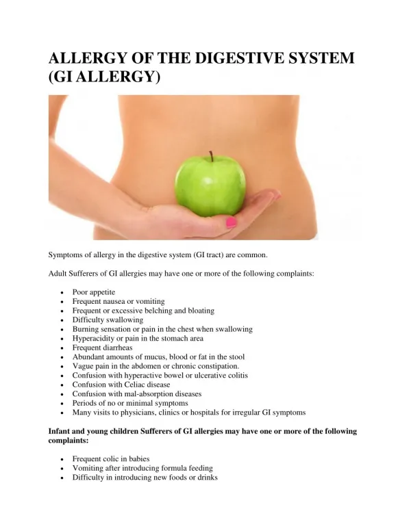
Download
1 / 28
340 likes | 766 Vues
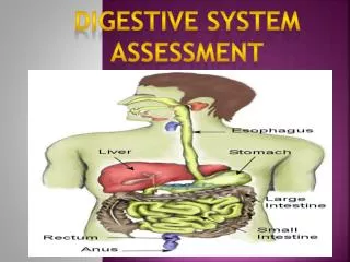
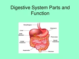
Assessment of Digestive and GI Function. Connie K. Cupples, MS, MSN, RN Union University. Outcome 1. Review the structure and function of the organs of the GI tract. A. Draw abdomen depicting organs in the four quadrants. B. Identify function of each of the organs of digestion.

E N D
Assessment of Digestive and GI Function Connie K. Cupples, MS, MSN, RN Union University
Outcome 1 • Review the structure and function of the organs of the GI tract. • A. Draw abdomen depicting organs in the four quadrants. • B. Identify function of each of the organs of digestion.
Function of Organs • Mouth • Stomach • Small Intestine • Colon
Outcome 2 • Explain the processes involved in the digestion, absorption, and elimination of food products. A. Identify major digestive enzymes, sources, and digestive actions. refer to pg. 943 table 34-1 Action of enzymes that digest CHO, protein, and fat
Outcome 3 • Describe assessment paramaters and techniques used when evaluating the GI tract. • A. Draw a torso and shade common sites of referred abdominal pain. • B. Discuss the order of the examination of the abdomen and give the rationale.
Health History • Focus on symptoms common to GI dysfunction: Pain Indigestion Intestinal gas Change in bowel habits Change in stool characteristics
Physical Assessment • Inspect mouth and contents • Supine with knees flexed slightly • Order of assessment: inspection auscultation palpation percussion Rationale for order of GI Assessment?
Outcome 4 • Describe preparation, education, and follow-up care for patients undergoing the following diagnostic testing of the GI tract. • A. Discuss how the nurse would prepare and educate patients for GI tests, including post procedure interventions.
Stool Tests • Collect on random basis except specimen where quantitative study is performed (fecal fat and urobilinogen) • Refrigerate quantitative specimen (24-72 hr. collections) • Special diet required for some tests • Fecal occult blood tests (foods & meds may alter results.
Abdominal Ultrasonography • Noninvasive means of imaging abdominal organs and structures. • NPO for 8-12 hrs. • Fat-free meal at supper if GB studies are done • Schedule barium studies after test if ordered as well
Upper GI X-ray Studies X-ray imaging after contrast media injested. Low residue diet X several days prior NPO after MN Laxative prep Discourage smoking day of test Hold all meds on day of test Monitor post test for 3 days to get rid of barium (fluids, laxatives, enemas)
Lower GI X-ray Studies • Visualization of lower GI tract after instillation of barium • Bowel prep to cleanse lower bowel • Low-residue diet 1-2 days prior • C/L diet for supper, laxative HS, NPO after MN • Cleansing enemas until clear in a.m. • Post procedure elimination of barium
CT • Cross-sectional images of abdominal organs and structures • NPO 6-8 hrs. before test • Question about contrast dye allergies • Schedule barium studies after CT scan if ordered
MRI • Noninvasive technique to supplement ultrasonography and CT scanning • NPO 6-8 hrs. prior to test • Remove all jewelry & other metals • Explain that procedure lasts 30-90 minutes • Explain type of equipment used (pt. may experience claustrophobia & hear knocking sound)
Esophagogastroduodenoscopy • Visualization of upper GI tract with fiberoptic lenses. • NPO 6-12 hrs. • Pt. may gargle with local anesthetic • Versed given IV – monitor pt. per conscious sedation protocol • Atropine may be given to dry secretions • Position on left side during procedure
Nursing Interventions Post EGD • Keep NPO until return of gag reflex • Simms position until awake, then semi-fowlers • Observe for s/s of perforation (pain, bleeding, unusual difficulty swallowing, temp) • Monitor for changes in P & BP • Instruct not to drive for 10-12 hrs. post
Colonsocopy • Direct visual inspection of the colon using flexible fiberoptic colonoscope. • Colon preparation a must • C/L diet at noon day before • Laxatives the night before • Use of preparation such as Golytely to lavage the bowel over 3-4 hrs. until returns are clear
Nursing Interventions post Colonoscopy • Monitor according to conscious sedation guidelines • Observe for s/s of bowel perforation (abdominal pain, distention, rectal bleeding, temp) • Provide written instructions due to amnesic effect of meds
Gastic Analysis, Gastic Acid Stimulation test & pH Monitoring • Determines secretory activity of gastic mucosa and gastric retention. • NPO 8-12 hrs. • Hold meds that affect gastic secretion • Smoking not allowed day of test • Insert small NG tube • Aspirate entire contents of stomach q 15 minutes for 1 hr.
Gastic Acid Stimulation • Med given to stimulate gastric secretion • Inform pt. that flushing may occur • Monitor P & BP q 15 min. • Collect gastic specimen q 15 minutes for 1 hr.
pH Monitoring • NPO 6 hrs. before test • Hold meds 24-36 hrs. • Probe inserted through nose • Connected to external recording device