Download
1 / 54
540 likes | 773 Vues
Digestive and Gastrointestinal(GI) Function. Miss Fatima Hirzallah. Objectives. On completion of this chapter, the learner will be able to: Describe the structure and function of the organs of the gastrointestinal (GI) tract..

E N D
Digestive and Gastrointestinal(GI) Function Miss Fatima Hirzallah
Objectives • On completion of this chapter, the learner will be able to: • Describe the structure and function of the organs of the gastrointestinal (GI) tract.. • Use assessment parameters appropriate for determining the status of GI function. • Describe the appropriate preparation, teaching, and follow-up care for patients who are undergoing diagnostic testing of the GI tract.
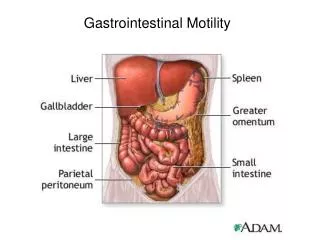
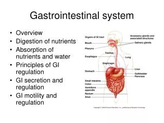
Anatomy of the GI Tract • The gastrointestinal (GI) tract is a 23- to 26-foot-long pathway that extends from the mouth to the esophagus, stomach, small and large intestines, and rectum, to the terminal structure, the anus .
An individual’s nutritional status depends not only on the type and amount of intake but also on the functioning of the gastric and intestinal portions of the gastrointestinal (GI) system.
Gastritis (inflammation of the gastric or stomach mucosa) is a common GI problem. Gastritis may be acute, lasting several hours to a few days, or chronic, resulting from repeated exposure to irritating agents or recurring episodes of acute gastritis. • Acute gastritis is often caused by dietary indiscretion—the person eats food that is contaminated with disease-causing microorganisms or that is irritating or too highly seasoned
Gastritis • Other causes of acute gastritis include overuse of aspirin and other nonsteroidal anti-inflammatory drugs (NSAIDs), • excessive alcohol intake, bile reflux, and radiation therapy. • Severe form of acute gastritis is caused by the ingestion of strong acid or alkali, which may cause the mucosa to become gangrenous or to perforate.
Scarring can occur, resulting in pyloric stenosis or obstruction. • Acute gastritis also may develop in acute illnesses, especially when the patient has had major traumatic injuries; burns; severe infection; hepatic, renal, or respiratory failure; or major surgery. Gastritis may be the first sign of an acute systemic infection.
Gastritis • Chronic gastritis and prolonged inflammation of the stomach may be caused by either benign or malignant ulcers of the stomach or by the bacteria Helicobacter pylori. • Chronic gastritis is sometimes associated with autoimmune diseases such as pernicious anemia; dietary factors such as caffeine; the use of medications, especially NSAIDs; alcohol; smoking; or reflux of intestinal contents into the stomach.
Pathophysiology • In gastritis, the gastric mucous membrane becomes edematous and hyperemic (congested with fluid and blood) and undergoes superficial erosion. • It secretes a scanty amount of gastric juice, containing very little acid but much mucus. Superficial ulceration may occur and can lead to hemorrhage.
Clinical Manifestations • The patient with acute gastritis may have abdominal discomfort, headache, lassitude, nausea, anorexia, vomiting, and hiccupping. • Some have no symptoms.
The patient with chronic gastritis may complain of anorexia, heartburn after eating,, a sour taste in the mouth, or nausea and vomiting. • Patients with chronic gastritis from vitamin deficiency usually have evidence of malabsorption of vitamin B12 caused by antibodies against intrinsic factor.
Assessment and Diagnostic Findings • Gastritis is sometimes associated with hypochlorhydria (absence or low levels of hydrochloric acid [HCl]) or with hyperchlorhydria (high levels of HCl).
Diagnosis can be determined by endoscopy, upper GI radiographic studies, and histological examination of a tissue specimen- biopsy. • diagnostic measures for detecting H. pylori include serologic testing for antibodies against the H. pylori antigen, and a breath test
Medical Management • The gastric mucosa is capable of repairing itself after a bout of gastritis. As a rule, the patient recovers in about 1 day. • nonirritating diet is recommended. • If bleeding is present, management is similar to the procedures used for upper GI tract hemorrhage • If it caused by ingestion of strong acids or alkalis, treatment consists of diluting and neutralizing the offending agent. To neutralize acids, common antacids (eg, aluminum hydroxide)
Medical Management • to neutralize an alkali, diluted lemon juice . • If corrosion is extensive or severe, emetics and lavage are avoided because of the danger of perforation and damage to the esophagus. • Therapy is supportive and may include nasogastric (NG) intubation .analgesic agents and sedatives, antacids, and intravenous (IV) fluids.
Fiberoptic endoscopy may be necessary. In extreme cases, emergency surgery may be required to remove gangrenous or perforated tissue.
A gastric resection or a gastrojejunostomy (anastomosis of jejunum to stomach to detour around the pylorus) may be necessary to treat pyloric obstruction, a narrowing of the pyloric orifice that cannot be relieved by medical management.
Medical Management • Chronic gastritis is managed by modifying the patient’s diet, promoting rest, reducing stress, and initiating pharmacotherapy. • H. pylori may be treated with antibiotics (eg, tetracycline or amoxicillin, combined with clarithromycin) and a proton pump inhibitor (eg, lansoprazole [Prevacid]), and possibly bismuth salts (Pepto-Bismol). • Research is being conducted to develop a vaccine against H. pylori
Gastric and Duodenal Ulcers • A peptic ulcer is an excavation (hollowed-out area) that forms in the mucosal wall of the stomach, in the pylorus (opening between stomach and duodenum), in the duodenum (first part of small intestine), or in the esophagus. • A peptic ulcer is frequently referred to as a gastric, duodenal, or esophageal ulcer, depending on its location, or as peptic ulcer disease.
Peptic ulcers are more likely to be in the duodenum than in the stomach. As a rule they occur alone, but they may occur in multiples. Chronic gastric ulcers tend to occur in the lesser curvature of the stomach, near the pylorus. • In the past, stress and anxiety were thought to be causes of ulcers. Research has identified that peptic ulcers result from infection with the gram-negative bacteria H. pylori
Familial tendency may be a significant predisposing factor. A further genetic link is noted in the finding that people with blood type O are more susceptible to peptic ulcers than are those with blood type A, B, or AB. There also is an association between duodenal ulcers and chronic pulmonary disease or chronic renal disease.
Stress ulcers, which are clinically different from peptic ulcers, are ulcerations in the mucosa that can occur in the gastroduodenal area. Stress ulcers may occur in patients who are exposed to stressful conditions.
Pathophysiology • Peptic ulcers occur mainly in the gastroduodenal mucosa because this tissue cannot withstand the digestive action of gastric acid (HCl) and pepsin. The erosion is caused by the increased concentration or activity of acid-pepsin, or by decreased resistance of the mucosa. • A damaged mucosa cannot secrete enough mucus to act as a barrier against HCl. • The use of NSAIDs inhibits the secretion of mucus that protects the mucosa. Patients with duodenal ulcer disease secrete more acid than normal, whereas patients with gastric ulcer tend to secrete normal or decreased levels of acid.
Pathophysiology • Stress ulcer is the term given to the acute mucosal ulceration of the duodenal or gastric area that occurs after physiologically stressful events, such as burns, shock, severe sepsis, and multiple organ traumas. • Differences of opinion exist as to the actual cause of mucosal ulceration in stress ulcers. Usually, it is preceded by shock; this leads to decreased gastric mucosal blood flow and to reflux of duodenal contents into the stomach. In addition, large quantities of pepsin are released. The combination of ischemia, acid, and pepsin creates an ideal climate for ulceration.
Pathophysiology • Stress ulcers should be distinguished from Cushing’s ulcers and Curling’s ulcers, two other types of gastric ulcers. • Cushing’s ulcers are common in patients with trauma to the brain. • Curling’s ulcer is frequently observed about 72 hours after extensive burns
Comparing Duodenal and Gastric Ulcers • DUODENAL ULCER Incidence • Age 30–60 • Male: female 2–3:1 • 80% of peptic ulcers are duodenal GASTRIC ULCER • Usually 50 and over • Male: female 1:1 • 15% of peptic ulcers are gastric
Signs, Symptoms, and Clinical Findings • DUODENAL ULCER • Hypersecretion of stomach acid (HCl) • May have weight gain • Pain occurs 2–3 hours after a meal; often awakened between 1–2 AM; • ingestion of food relieves pain • Vomiting uncommon • GASTRIC ULCER • Normal—hyposecretion of stomach acid (HCl) • Weight loss may occur • Pain occurs 1⁄2 to 1 hour after a meal; rarely occurs at night; may be relieved by vomiting; • ingestion of food does not help, sometimes increases pain • Vomiting common
Comparing Duodenal and Gastric Ulcers • DUODENAL ULCER • Hemorrhage less likely than with gastric ulcer, but if present melena more common than Hematemesis More likely to perforate than gastric ulcers • GASTRIC ULCER • Hemorrhage more likely to occur than with duodenal ulcer; hematemesis more common than melena
Comparing Duodenal and Gastric Ulcers • DUODENAL ULCER • Malignancy Possibility • Rare • Risk Factors • H. pylori, alcohol, smoking, cirrhosis, stress • GASTRIC ULCER • Occasionally • H. pylori, gastritis, alcohol, smoking, use of NSAIDs, stress
Clinical manifestation • Symptoms of an ulcer may last for a few days, weeks, or months and may disappear only to reappear, often without an identifiable cause. Many people with ulcers have no symptoms, and perforation or hemorrhage may occur in 20% to 30% of patients who had no preceding manifestations.
Clinical Manifestations • As a rule, the patient with an ulcer complains of dull, pain or a burning sensation in the midepigastrium or in the back. It is believed that the pain occurs when the increased acid content of the stomach and duodenum erodes the lesion and stimulates the exposed nerve endings.
pyrosis (heartburn), vomiting, constipation or diarrhea, and bleeding. Pyrosis is a burning sensation in the esophagus and stomach that moves up to the mouth. Heartburn is often accompanied by sour taste , or burping, which is common when the patient’s stomach is empty. • Fifteen percent of patients with gastric ulcers experience bleeding.
Assessment and Diagnostic Findings • A physical examination may reveal pain, epigastric tenderness, or abdominal distention. • A barium study of the upper GI tract may show an ulcer; however, endoscopy is the preferred diagnostic procedure because it allows direct visualization of inflammatory changes, ulcers, and lesions-biopsy.
Stools may be tested periodically until they are negative for occult blood. Gastric secretory studies are of value in diagnosing achlorhydria • pylori infection may be determined by biopsy and histology with culture. • There is also a breath test that detects H. pylori, as well as a serologic test for antibodies to the H. pylori antigen.
Medical Management • peptic ulcers treated with antibiotics to eradicate H. pylori have a lower recurrence rate than those not treated with antibiotics. The goals are to eradicate H. pylori and to manage gastric acidity. Methods used include medications, lifestyle changes, and surgical intervention.
Pharmacologic Therapy • Currently, the most commonly used therapy for peptic ulcers is a combination of antibiotics, proton pump inhibitors, and bismuth salts that suppress or eradicate H. pylori.
Histamine-2 (H2) receptor antagonists and proton pump inhibitors are used to treat NSAID-induced ulcers and other ulcers not associated with H. pylori infection.
Nursing Diagnoses • Acute pain related to irritated stomach mucosa • Imbalanced nutrition, less than body requirements, related to inadequate intake of nutrients • Risk for imbalanced fluid volume related to insufficient fluid intake and excessive fluid loss subsequent to vomiting • Anxiety related to treatment
Surgical Management • The introduction of antibiotics to eradicate H. pylori and of H2 receptor antagonists as treatment for ulcers has greatly reduced the need for surgical intervention. However, surgery is usually recommended for patients with intractable ulcers (those that fail to heal after 12 to 16 weeks of medical treatment), life-threatening hemorrhage, perforation.
obstruction and for those with hypersecreation of acid not responding to medications . • Surgical procedures include vagotomy , Antrectomy (Gastrojejunostomy), Gastroduodenostomy)
vagotomy • Decreases gastric acid by diminishing cholinergic stimulation to the parietal cells, making them less responsive to gastrin. May be performed via open surgical approach, laparoscopy, or thoracoscopy
(Gastroduodenostomy • Removal of the lower portion of the antrum of the stomach (which contains the cells that secrete gastrin)as well as a small portion of the duodenum
(Gastrojejunostomy) : Removal of lower portion (antrum) of stomach with anastomosis to jejunum • Complication :Dumping syndrome, anemia, mal-absorption, weight loss. Recurrence rate of ulcer is 10%–15%