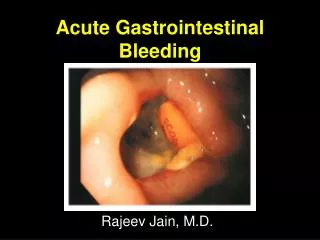
Gastrointestinal Bleeding
Gastrointestinal Bleeding. PCOM Internal Medicine Residents 2004. GI Bleeding. Initial Evaluation Approach to the Patient Sources Upper GI Bleeds Lower GI Bleeds Etiology Management Admission Orders. Initial Evaluation. History and Physical points to Source/Etiology

Gastrointestinal Bleeding
E N D
Presentation Transcript
Gastrointestinal Bleeding PCOM Internal Medicine Residents 2004
GI Bleeding • Initial Evaluation • Approach to the Patient • Sources • Upper GI Bleeds • Lower GI Bleeds • Etiology • Management • Admission Orders
Initial Evaluation • History and Physical points to Source/Etiology • History of Present Illness • Attention to PMHx, Social Hx, Medications
History • Hematemesis (coffee grounds vs. bright red) • Hematochezia • Melena - dark, tarry stool • Pain symptoms • Medications – NSAIDs, steroids, ASA, Plavix, Coumadin, Lovenox, Heparin, Iron • PMHx - arthritis, ulcer disease, EtOH
Good Thorough Physical Exam Including: • HR, BP, tilt test, RR, O2 saturation • General appearance, Mental status • Neck veins, oral mucosa • Skin temperature and color • Abdominal exam • Rectal • Stigma of Cirrhosis • NG Tube findings (upper vs. lower g.i. source) • Urine output
Approach to the patient • Labs • CBC • Serial HgB • Platelets • BMP • BUN, Cr • Type and Crossmatch • Coagulation studies • Imaging studies?
Sources of GI Bleeding • Upper GI Tract • Proximal to the Ligament of Treitz • 70% of GI Bleeds • Lower GI Tract • Distal to the Ligament of Treitz • 30% of GI Bleeds
Localization of Bleeding • History • NG Tube • EGD • Colonoscopy • Tagged RBC Scan • Angiography
Upper GI Bleed • 50% present with hematemesis • NGT with positive blood on aspirate • 11% of brisk bleeds have hematochezia • Melena (black tarry stools)—this develops with apporximately 150-200cc of blood in the upper GI tract. • Stool turns black after 8 hours of sitting within the gut.
Upper GI Bleed • Risk Factors • NSAID use • H. pylori infection • Increased age • Upper GI Bleeding accounts for approximately 350,000 hospitalizations per year.
Upper GI Bleed • Etiology of Upper Bleeds • Duodenal Ulcer-30% • Gastric Ulcer-20% • Varices-10% • Gastritis and duodenitis-5-10% • Esophagitis-5% • Mallory Weiss Tear-3% • GI Malignancy-1% • Dieulafoy Lesion • AV Malformation-angiodysplasia
GI Malignancy • Esophageal Tumor
GI Malignancy • Gastric Carcinoma
Lower GI Bleed • Hematochezia • Blood in Toilet • Clear NGT aspirate • Normal Renal Function • Usually Hemodynamically stable Only 1/3 of patients with lower GI bleeds have positive orthostatics (tilt test).
Lower GI Bleed • Etiology of Lower Bleeds • Diverticular-20% • AVM-10% • Malignancy-2-26% • Inflammatory Bowel Disease-10% • Ischemic Colitis • Acute Infectious Colitis • Radiation Colitis/Proctitis • Aortoenteric Fistula
Malignancy • Colon Carcinoma
Management of GI Bleed • Oxygen • IV Access-central line or two large bore peripheral IV sites • Isotonic saline for volume resuscitation • Start transfusing blood products if the patient remains unstable despite fluid boluses. • Airway Protection • Altered Mental Status and increased risk of aspiration with massive upper GI bleed.
Management of GI Bleed • ICU admit indications • Significant bleeding with hemodynamic instability • Transfusion • Brisk Bleed, transfusing should be based on hemodynamic status, not lab value of Hgb. • Cardiopulmonary symptoms-cardiac ischemia or shortness of breath, decreased pulse ox • 1 unit PRBC increases Hgb by 1mg/dL and increase Hct by 3% • FFP for INR greater than 1.5 • Platelets for platelet count less than 50K
Basic Admission Orders • Admit to ICU/intermediate care/telemetry s/o … • Dx: Upper/Lower G.I. Bleed • Condition: • VS: • Allergies: • Activity: Bedrest • Nursing: Is/Os, ? Foley • Diet: NPO
Basic Admission Orders (Cont.) • IVF: NSS @ ?cc/h • Medications: I.V. Protonix, convert medications to i.v., hold anti-hypertensives • Labs: serial H/H, type and cross, coags, Chem 7, LFTs • Consults: g.i., surgery?
References • Harrison’s Principles of Internal Medicine 14th edition • Gastrointestinal Atlas.com endoscopy photos