
The Digestive system
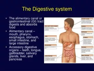
The Digestive system. The alimentary canal or gastrointestinal (GI) tract digests and absorbs food Alimentary canal – mouth, pharynx, esophagus, stomach, small intestine, and large intestine Accessory digestive organs – teeth, tongue, gallbladder, salivary glands, liver, and pancreas.

The Digestive system
E N D
Presentation Transcript
The Digestive system • The alimentary canal or gastrointestinal (GI) tract digests and absorbs food • Alimentary canal – mouth, pharynx, esophagus, stomach, small intestine, and large intestine • Accessory digestive organs – teeth, tongue, gallbladder, salivary glands, liver, and pancreas
Organization of the Digestive tract • Mucosa lines digestive tract (mucous epithelium) • Moistened by glandular secretions • Lamina propria and epithelium form mucosa • Submucosa - layer of dense irregular connective tissue • Muscularis externa - smooth muscle arranged in circular and longitudinal layers • Serosa - serous membrane covering most of the muscularis externa
Digestive System Anatomy Figure 21-2c
Digestive System Activities • The GI tract is a “disassembly” line • Nutrients become more available to the body in each step • Ingestion – taking food into the digestive tract • Mechanical digestion – chewing, churning food, segmentation • Propulsion – swallowing and peristalsis • Chemical digestion – catabolic breakdown of food • Absorption – movement of nutrients from the GI tract to the blood or lymph • Excretion – elimination of indigestible solid wastes
Basic Processes of the Digestive System Figure 21-1
Motility • Movement of digestive materials • Visceral smooth muscle • Tonic contractions • Sustained • Smooth muscle sphincters and stomach • Phasic contractions • Rhythmic cycles of activity - pacemaker cells • Last a few seconds • Peristalsis moves bolus forward • Segmentation mixes
Autonomous Smooth Muscle • Pacesetter cells • Slow wave potentials – digestive tracts’s basic electrical rhythm • Wavelike fluctuations in membrane potential • Transmitted throughout smooth muscle via gap junctions • Threshold is reached by the effect of various mechanical, neural and hormonal factors
Motility • Peristalsis - waves that move a bolus
Motility • Segmentation – a churn and fragment a bolus
Ingestion and Mechanical Digestion • Food is ingested • Mechanical digestion begins (chewing) • Propulsion is initiated by swallowing • Salivary amylase begins chemical breakdown of starch • The pharynx and esophagus serve as conduits to pass food from the mouth to the stomach • Uvula guards opening to pharynx
The Oral Cavity • Mechanical processing by the teeth, tongue, and palatal surfaces • Lubrication • Assistance in swallowing • Limited digestion Figure 24.6a, b
Salivary glands (three pairs) • Parotid, sublingual, and submandibular glands produce saliva • Stimulated by thought of food or ingested food • Secreted from serous and mucous cells of salivary glands • Watery solution includes electrolytes, buffers, glycoproteins, antibodies, enzymes • Functions include: • Lubrication, moistening, and dissolving • Initiation of digestion of complex carbohydrates • Strong sympathetic stimulation inhibits salivation and results in dry mouth
Swallowing (deglutition) Process • Involves the coordinated activity of the tongue, soft palate, pharynx, esophagus and 22 separate muscle groups • Three phases • Buccal phase – tongue pushes bolus against soft palate and forced into the oropharynx, triggering swallowing reflex – controlled by the medulla and lower pons • Pharyngeal – esophogeal sphincter relaxes while epiglottis closes • Esophageal – bolus moves into esophagus propelled by peristalsis and into stomach Figure 24.11a-h
Functions of the stomach • Holds ingested food • Degrades this food both physically and chemically • Delivers chyme to the small intestine • Enzymatically digests proteins with pepsin • Secretes intrinsic factor required for absorption of vitamin B12
Stomach Lining • The stomach is exposed to the harshest conditions in the digestive tract • To keep from digesting itself, the stomach has a mucosal barrier with: • A thick coat of bicarbonate-rich mucus on the stomach wall • Epithelial cells that are joined by tight junctions • Gastric glands that have cells impermeable to HCl
Glands of the Stomach • Gastric glands have a variety of secretory cells • Mucous neck cells – mucus • Parietal cells – HCl and intrinsic factor • Chief cells – pepsinogen • Pepsinogen is activated to pepsin by HCl in the stomach • Pepsin itself via a positive feedback mechanism • Enteroendocrine cells – gastrin, histamine, cholecystokinin (CCK
Regulation of Gastric Secretion • Neural and hormonal mechanisms regulate the release of gastric juice • Stimulatory and inhibitory events occur in three phases • Cephalic (reflex) phase: prior to food entry • Gastric phase: once food enters the stomach • Intestinal phase: as partially digested food enters the duodenum
Cephalic Phase • Cephalic phase prepares stomach to receive ingested material • Directed by CNS via the vagus nerve • Stimulated by sight, smell, taste, or thought of food • Accelerates gastric juices • Inhibited by loss of appetite, depression
Gastric Phase • Enhanced secretion of gastric juices due to the arrival of food in the stomach • Homogenize and acidify chyme • Production of pepsinogen - digestion of proteins • Stimulated by • Stomach distension - activation of stretch receptors • Chemoreceptors detects - peptides, caffeine, and rising pH • Neural - plexuses • Hormonal - secretion of gastrin • Inhibitory events include: • A pH lower than 2 • Emotional upset that overrides the parasympathetic division
Intestinal phase • Intestinal phase – release of hormones controls the rate of gastric emptying • Excitatory phase – distension of duodenum, presence of partially digested foods • Releases enterogastrones that inhibit gastric secretion: CCK, GIP, Secretin • Inhibited by low pH, presence of fatty, acidic, or hypertonic chyme, and/or irritants in the duodenum
Release of Gastric Juice Figure 23.16
Regulation of HCl Secretion • HCl secretion is stimulated by ACh, histamine, and gastrin through second-messenger systems • Release of hydrochloric acid: • Is low if only one ligand binds to parietal cells • Is high if all three ligands bind to parietal cells • Antihistamines block H2 receptors and decrease HCl release
Gastric Contractile Activity • Peristaltic waves move toward the pylorus at the rate of 3 per minute • This basic electrical rhythm (BER) is initiated by pacemaker cells • Most vigorous peristalsis and mixing occurs near the pylorus • Chyme is either: • Delivered in small amounts to the duodenum or • Forced backward into the stomach for further mixing
Regulation of Gastric Emptying • Gastric emptying is regulated by: • The neural enterogastric reflex • Hormonal (enterogastrone) mechanisms • These mechanisms inhibit gastric secretion and duodenal filling • Carbohydrate-rich chyme quickly moves through the duodenum • Fat-laden chyme is digested more slowly causing food to remain in the stomach longer
Gastrointestinal Hormones • Gastrin • Release is stimulated by presence of protein in stomach • Secretion inhibited by accumulation of acid in stomach • Acts in several ways to increase secretion of HCl and pepsinogen • Enhances gastric motility, stimulates ileal motility, relaxes ileocecal sphincter, induces mass movements in colon • Helps maintain well-developed, functionally viable digestive tract lining
Gastrointestinal Hormones • Secretin • Presence of acid in duodenum stimulates release • Inhibits gastric emptying in order to prevent further acid from entering duodenum until acid already present is neutralized • Inhibits gastric secretion to reduce amount of acid being produced • Stimulates pancreatic duct cells to produce large volume of aqueous NaHCO3 secretion • Stimulates liver to secrete NaCO3 rich bile which assists in neutralization process • Along with CCK, is trophic to exocrine pancreas
Gastrointestinal Hormones • CCK • Inhibits gastric motility and secretion • Stimulates pancreatic acinar cells to increase secretion of pancreatic enzymes • Causes contraction of gallbladder • Along with secretin, is trophic to exocrine pancreas • Implicated in long-term adaptive changes in proportion of pancreatic enzymes in response to prolonged diet changes • Important regulator of food intake • GIP • Glucose-dependent insulinotrophic peptide • Stimulates insulin release by pancreas
Regulation of Gastric Emptying Figure 23.19
Digestion And Absorption In The Stomach • Preliminary digestion of proteins - pepsin • Permits digestion of carbohydrates • Very little absorption of nutrients • Some drugs, however, are absorbed
Small intestine • Important digestive and absorptive functions • Secretions and buffers provided by pancreas, liver, gall bladder • Three subdivisions: • Duodenum • Jejunum • Ileum • Ileocecal sphincter - transition between small and large intestine
Small Intestine • Structural modifications of the small intestine wall increase surface area • Plicae circulares: deep circular folds of the mucosa and submucosa • Villi – fingerlike extensions of the mucosa • Microvilli – tiny projections of absorptive mucosal cells’ plasma membranes
Small Intestine • The epithelium of the mucosa is made up of: • Absorptive cells and goblet cells • Enteroendocrine cells • Interspersed T cells called intraepithelial lymphocytes (IELs) • Cells of intestinal crypts secrete intestinal juice • Secreted in response to distension or irritation of the mucosa • Slightly alkaline and isotonic with blood plasma • Largely water, enzyme-poor, but contains mucus
Small Intestine • Glands of the duodenum • Moisten chyme • Help buffer acids • Maintain digestive material in solution • Hormones • Secretin - produces alkaline buffers, increase bile by liver and pancreas • Cholecystokinin – increase pancreatic enzymes, stimulates contraction of gall bladder, reduces hunger sensation • GIP – stimulates release of insulin, inhibits gastric secretion and motility
Activities of Major Digestive Tract Hormones Figure 24.22
The liver • The largest gland in the body • Performs metabolic and hematological regulation and produces bile • Histological organization • Lobules containing single-cell thick plates of hepatocytes • Lobules unite to form common hepatic duct • Duct meets cystic duct to form common bile duct
The Liver • Hexagonal-shaped liver lobules are the structural and functional units of the liver • Composed of hepatocytes • Hepatocytes’ functions include: • Production of bile • Processing bloodborne nutrients • Storage of fat-soluble vitamins • Detoxification • Secreted bile flows between hepatocytes toward the bile ducts in the portal triads
Composition of Bile • A yellow-green, alkaline solution containing bile salts, bile pigments, cholesterol, neutral fats, phospholipids, and electrolytes • Bile salts are cholesterol derivatives that: • Emulsify fat • Facilitate fat and cholesterol absorption • Help solubilize cholesterol • Enterohepatic circulation recycles bile salts • The chief bile pigment is bilirubin, a waste product of heme
The Gallbladder • Thin-walled, green muscular sac on the ventral surface of the liver • Stores and concentrates bile by absorbing its water and ions • Releases bile via the cystic duct, which flows into the bile duct
Regulation of Bile Release • Acidic, fatty chyme causes the duodenum to release: • Cholecystokinin (CCK) and secretin into the bloodstream • Bile salts and secretin transported in blood stimulate the liver to produce bile • Vagal stimulation causes weak contractions of the gallbladder • Cholecystokinin causes: • The gallbladder to contract • The hepatopancreatic sphincter to relax • As a result, bile enters the duodenum
Regulation of Bile Release Figure 23.25
The Pancreas • Pancreatic duct penetrates duodenal wall • Endocrine functions - insulin and glucagons • Exocrine functions • Secretes pancreatic juice secreted into small intestine which breaks down all categories of foodstuff • Acini (clusters of secretory cells) contain zymogen granules with digestive enzymes
Composition of Pancreatic Juice • Water solution of enzymes and electrolytes (primarily HCO3–) • Neutralizes acid chyme • Provides optimal environment for pancreatic enzymes • Enzymes are released in inactive form and activated in the duodenum • Examples include • Trypsinogen is activated to trypsin • Procarboxypeptidase is activated to carboxypeptidase • Active enzymes secreted • Amylase, lipases, and nucleases • These enzymes require ions or bile for optimal activity
Regulation of Pancreatic Secretion • Secretin and CCK are released when fatty or acidic chyme enters the duodenum • CCK and secretin enter the bloodstream • Upon reaching the pancreas: • CCK induces the secretion of enzyme-rich pancreatic juice • Secretin causes secretion of bicarbonate-rich pancreatic juice • Vagal stimulation also causes release of pancreatic juice
Regulation of Pancreatic Secretion Figure 23.28
Digestion in the Small Intestine • As chyme enters the duodenum: • Carbohydrates and proteins are only partially digested • No fat digestion has taken place • Digestion continues in the small intestine • Chyme is released slowly into the duodenum • Because it is hypertonic and has low pH, mixing is required for proper digestion • Required substances needed are supplied by the liver • Virtually all nutrient absorption takes place in the small intestine
Motility in the Small Intestine • The most common motion of the small intestine is segmentation • It is initiated by intrinsic pacemaker cells (Cajal cells) • Moves contents steadily toward the ileocecal valve • After nutrients have been absorbed: • Peristalsis begins with each wave starting distal to the previous • Meal remnants, bacteria, mucosal cells, and debris are moved into the large intestine
Control of Motility • Local enteric neurons of the GI tract coordinate intestinal motility • Cholinergic neurons cause: • Contraction and shortening of the circular muscle layer • Shortening of longitudinal muscle • Distension of the intestine • Other impulses relax the circular muscle • The gastroileal reflex and gastrin: • Relax the ileocecal sphincter • Allow chyme to pass into the large intestine
Functions of the large intestine • Reabsorb water and compact material into feces • Absorb vitamins produced by bacteria • Store fecal matter prior to defecation
Functions of the Large Intestine • Other than digestion of enteric bacteria, no further digestion takes place • Vitamins, water, and electrolytes are reclaimed • Its major function is propulsion of fecal material toward the anus • Though essential for comfort, the colon is not essential for life


