
RESPIRATORY SYSTEM
Explore the key functions of the respiratory system - from respiration to sound production and ventilation. Learn about the terms related to nasal cavity, sinuses, larynx, and more. Enhance your knowledge!

RESPIRATORY SYSTEM
E N D
Presentation Transcript
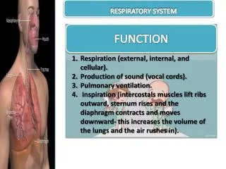
RESPIRATORY SYSTEM FUNCTION Respiration (external, internal, and cellular). Production of sound (vocal cords). Pulmonary ventilation. Inspiration (intercostals muscles lift ribs outward, sternum rises and the diaphragm contracts and moves downward- this increases the volume of the lungs and the air rushes in).
TERMS TO REMEMBER • nasal cavity - Contains nasal septum, turbinate, and cilia. • nasal septum - Divides nasal cavities into right and left sides. • turbinate's - Bones that protrude into the nasal cavity- they increase surface area for filtering dust and dirt particles by the mucous membrane. • Cilia- Nosehairs, traplargerdirtparticles. • sinuses -Cavities in the skull, ducts connect them to the nasal cavity, lined with mucous membrane to warm and moisten the air. Give resonance to voice.
types of sinuses - Frontal, maxillary, ethmoid, and sphenoid. • pharynx - Throat. Common passageway for air and food. 5" long. • epiglottis - When food is swallowed, this closes over the opening to the larynx, preventing food from entering the lungs • larynx- Voice box. Triangular chamber below pharynx. "Adam's Apple". • Glottis- Vocal cords within the larynx. Trachea- Windpipe. 4.5" long. Walls are alternate bands of membrane and c-shaped rings of hyaline cartilage to keep it open. Lined with ciliated mucous membrane. Coughing and expectoration gets rid of dust-laden mucous.
Bronchi - Similar to trachea with ciliated mucous membrane and hyaline cartilage. Lower end of trachea divides into right and left this. • bronchial tubes - Cartilaginous plates (instead of c-shaped rings of trachea).. • Bronchioles - Thinner walls of smooth muscle, lined with ciliated epithelium. Subdivision of bronchi. At the end, alveolar duct and cluster of alveoli. • Alveoli -Composed of single layer of epithelial tissue. Inner surfaces covered with surfactant to keep from collapsing. Each surrounded by capillaries. Oxygen and carbon dioxide exchange takes place between these and capillaries.
lungs - Fill thoracic cavity. Tissue is porous and spongy- it floats. • Apex- Upper part of lung. • Base- Lower part of lung. • right lung- Larger and shorter (displaced by liver) and has three lobes. • left lung - Smaller (displaced by heart) and has two lobes. • Pleura - Thin, moist, slippery membrane that covers lungs. Double-walled sac. Space is pleural cavity- filled with pleural fluid to prevent friction. • coughing - Deep breath followed by forceful expulsion of air to clean lower respiratory tract. • hiccups - Spasm of diaphragm and spasmotic closure of the glottis- irritation to diaphragm or phrenic nerve. • Sneezing- Air forced through nose to clear respiratory tract. • yawning - Deep prolonged breath that fills the lungs, increases oxygen within the blood. • neural factors of breathing control - Respiratory center located in medulla oblongata (in the brain). Increase in CO2 and decrease in O2 in the blood will trigger respiratory center. • phrenic nerve- Stimulates the diaphragm.


