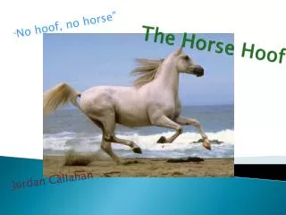
The Hoof
The Hoof. Stashak. Equine thoracic limb. Banks. Equine foot. Banks. Equine foot. Hoof - Epidermis. Wall Bars Sole Frog. Stashak Fig. 1.4. Hoof Epidermis (orange) Dermis (green). Bacha & Bacha Fig. 12.71. Hoof - Epidermis. Stashak Fig. 1.4. Hoof - Epidermis. Hoof wall Bars

The Hoof
E N D
Presentation Transcript
Stashak Equine thoracic limb
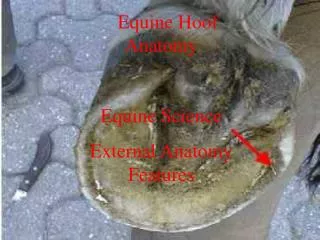
Banks Equine foot
Banks Equine foot
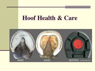
Hoof - Epidermis • Wall • Bars • Sole • Frog Stashak Fig. 1.4
Bacha & Bacha Fig. 12.71 Hoof - Epidermis Stashak Fig. 1.4
Hoof - Epidermis • Hoof wall • Bars • Sole • Frog
Hoof wall • Perioplic epidermis = stratum externum, stratum tectorium • Coronary epidermis = stratum medium, tubular epidermis • Tubular and intertubular horn • Laminar epidermis = stratum lamellatum • Non-tubular horn • Fuses to stratum medium and connects wall to dermis
epidermis Hoof Figure 34. Junction of epidermis and hoof, 6X. The figure shows the area of the periople; this is the region of the epidermis that gives rise to the stratum externum. Distal to the periople is the coronary region; this is the region where the stratum germinativum of the epidermis gives rise to the tubular and intertubular horns of the stratum medium. The coronary region is seen at higher magnifications in the figure that follows. periople stratum externum epidermis at coronary region coronary corium
Hoof wall • Perioplic epidermis, stratum externum • Coronary epidermis, stratum medium • Laminar epidermis, stratum lamellatum Stashak Fig. 1.5
Coronary epidermis, stratum medium • Tubular horn • Intertubular horn
Hoof stratum externum stratum medium stratum internum dermis (corium) Figure 28. Cross section of the wall of equine hoof, 6X. Section was taken from a young horse. The hoof is the horny epidermal covering of the distal end of the digit; it is composed of dead keratinized cells. The wall of the hoof, on cross section, shows a stratum externum (external layer), a stratum medium (middle layer), and a stratum internum (internal layer). An area similar to that in the inset is seen at higher magnifications in the figures that follow.
Hoof stratum externum stratum medium tubular horn intertubular horn Figure 29. Equine hoof, cross section. 20X. The figure shows the stratum externum and stratum medium. The stratum externum (stratum tectorium) is a thin layer of flat keratinized cells. The stratum medium comprises the bulk of the wall and is composed of tubular horn and intertubular horn. The inset is seen at higher magnifications in the next figures.
A Hoof tubular horn Figure 30 A-B. Stratum medium of hoof wall. A-50X, B-200X. The stratum medium is composed of tubular and intertubular horns. The inset in Figure A shows a tubular horn which is magnified in Figure B. The tubular horn is composed of spirally arranged cells that do not cornify (keratinize) as completely as those of the intertubular horn and they stain more intensely (basophilic). The intertubular horn is composed of flat keratinized layers of cells. intertubular horn B
Stratum lamellatum • Primary laminae • Secondary lamina
Hoof stratum externum stratum medium stratum internum dermis (corium) Figure 28. Cross section of the wall of equine hoof, 6X. Section was taken from a young horse. The hoof is the horny epidermal covering of the distal end of the digit; it is composed of dead keratinized cells. The wall of the hoof, on cross section, shows a stratum externum (external layer), a stratum medium (middle layer), and a stratum internum (internal layer). An area similar to that in the inset is seen at higher magnifications in the figures that follow.
Hoof primary laminae secondary laminae laminar corium Figure 31. Stratum internum of hoof wall. 20X. The stratum internum (stratum lamellatum) is composed of several primary laminae, each of which bears several secondary laminae. The vascular connective tissue between the laminae is the laminar corium (dermis).
A primary laminae Hoof Figure 32 A-B. Stratum internum of hoof wall. A-50X, B-200X. Figure A shows primary laminae with several secondary laminae. The laminae are seen at high magnification in Figure B. The primary lamina is composed of dead keratinized cells. The secondary laminae are composed of living cells of the stratum germinativum of the epidermis. secondary laminae blood vessel laminar corium primary lamina B secondary laminae blood vessel
Epidermis • Bars, sole, frog • Epidermis is similar to tubular regions of wall • Horn material is softer than wall
P1 Hoof DDF epidermis epidermis P2 dermis dermis Figure 37. Sagittal section of equine foot. 2X. This is the same section in Figure 33 and is being shown for orientation purpose. The figures that follow will show the region of the hoof sole (inset). ds coronary corium DC P3 (Stratum germinativum of epidermis is located here) wall of hoof sole of hoof
Hoof A B dermal papillae osseous tissue of P3 sole corium sole epidermis sole of hoof Figure 38A-B. Region of the sole of hoof. A-6X, B-20X. The structure of the sole is similar to that seen in the coronary region. Notice the dermal papilla extending downward into the epidermis of the sole. The epidermal cells will produce tubular and intertubular horns similar to those seen in the stratum medium of the hoof wall.
Dermis (corium) • Perioplic corium • Papillae are perpendicular to ground • Nourishes stratum externum • Coronary corium • Papillae are perpendicular to ground • Nourishes stratum medium • Laminar corium • Primary and secondary laminae instead of papillae • Laminae are perpendicular to ground • Nourishes stratum lamellatum
Corium of the Foot • Corium • Continuation of the skin • Contains elastic fibers and extensive venous plexus • Intimately associated with periosteum of bones in foot • Frog corium contains modified merocrine sweat glands • Digital cushion • Fibroelastic connective tissue • Acts as a shock absorber
Laminae • Sensitive lamina • Secondary epidermal lamina with nerve endings and germinal epithelium • Primary and secondary laminar corium • Insensitive lamina • Primary epidermal lamina – cornified material
Dog Claw Banks
Dog Claw Dellman Fig. 16.41
Dog Claw Aughey & Frye
Dog Claw Aughey & Frye