Download
1 / 20
210 likes | 500 Vues
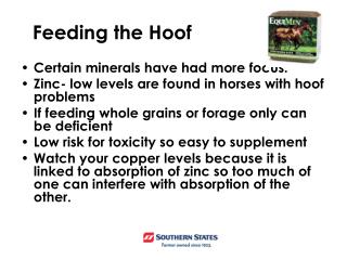
The Equine Hoof. By: Nadja Koehler. Hoof Wall. Made up of keratinized epithelial cells Cells are arranged in tubules and run from coronary band to ground surface Thickest at toe becoming thinner at quarters (sides) Contains pigment. Hoof Wall. Function: Weight-bearing surface of the hoof

E N D
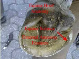
The Equine Hoof By: Nadja Koehler
Hoof Wall • Made up of keratinized epithelial cells • Cells are arranged in tubules and run from coronary band to ground surface • Thickest at toe becoming thinner at quarters (sides) • Contains pigment
Hoof Wall • Function: • Weight-bearing surface of the hoof • Helps retain moisture • Protect internal structures of the foot
Laminae • Two Layers: • Insensitive-forms inner layer of hoof wall • Sensitive-covers surface of coffin bone, acts as attachment for hoof wall and coffin bone, and acts as main area of circulation within foot • White Line-yellowish area where layers intermesh
Bars • Where hoof wall is reflected back toward toe • Located in heel area of hoof • Function: • Prevent over-expansion of hoof wall
Sole • Covers bottom of coffin bone • Sensitive • Self-limiting growth • Sloughs off when thickness > hoof wall • Concave at ground surface • Shape prevents sole from directly bearing weight • Easily bruised • Occurs when bearing weight: heavy riders, “flat feet”
Frog • Occupies area between bars • Wedge-shaped • Apex — point of frog • Cleft—ridge in rear portion of frog • Sensitive • Produced by papillae • Elastic • Moisture content ~ 50% • Greasy secretions from fat glands bet. digital cushion and frog
Digital Cushion • Also called plantar cushion • Fleshy “heel” • Back half of hoof • Fibro-elastic, fatty • Functions: • Shock absorber for foot • Pumps blood from foot back to heart
Bones • Three Bones: • Short pastern • Partly in and partly above hoof • Navicular bone • Smallest bone • Increases articular surface and movement of coffin bone
Bones Continued • Coffin bone • Location-to the front and slightly to outer side of hoof • Largest bone • Provides shape to foot and rigidity needed for weight-bearing • Resembles miniature hoof in shape
The Horse’s Second Heart What do I mean?? The Hoof, of course!
How? • Blood is pumped to the hoof from the heart through arteries • With each step, pressure is put on the veins in the plantar cushion of hoof which pumps the blood back to the heart • As this pressure is released, the blood flows back to the hoof through the arteries by a combination of heart pulses and gravity
Lameness • Most lameness can be prevented • How? • Proper foot care and management
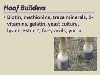
Healthy Hooves • Frog is a good indicator of foot health • Daily maintenance prevents lameness • Good foot care should include: • Regularity--Routine cleaning • Frequency--Periodic trimming • Cleanliness • Use of proper corrective measures--Corrections and treatment
Routine Cleaning • Includes use of: • Hoof pick • Fine-bristled wire brush • Always clean from heel toward toe • Do not apply too much pressure with either tool. This can cause: • Damage-bruising, abcess, infection, etc. • Disturbance of moisture balance
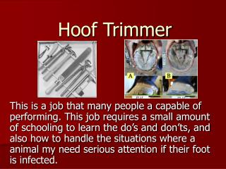
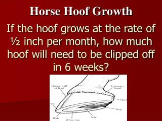
Trimming • Goal: • To maintain proper shape and length of the hoof • Hooves should be trimmed every 4-6 weeks depending on usage of your horse • Tools: • Hoof knife • Nippers • Rasp
What causes lameness? • Stone in the foot- • Stones lodge between shoe and frog • Bruised sole- • Direct injury of flat of foot by stones or irregular ground • Corns- • Bruising of sole between bar and hoof wall • Caused from poorly fitted shoes or neglect to reshod regularly
Causes continued... • Pricked foot or Puncture wounds- • Result from foreign objects entering sole (stone, glass, wire, etc.) • Foreign objects can stay in foot for as long as a year • Hoof cracks- • Occur mostly in dry or untrimmed hooves • Can also be caused by injury of hoof forming tissue • Thrush- • Bacterial infection of frog and sole due to irregular cleaning and dirty conditions
Causes continued... • Laminitis- • Inflammation of laminae • Caused by overeating of grain, ingestion of cold water by a hot horse, retained afterbirth, overfatness, idle horse on a lush pasture • Navicular disease- • Caused by injury to navicular bone • Common in breeds with genetic defects in conformation • Increased probability with heavy use on hard ground