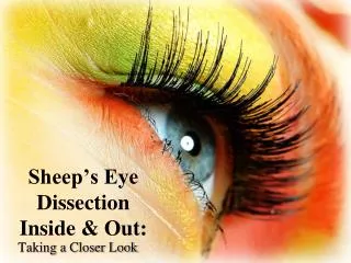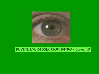

Eye Dissection Exploration Guide
E N D
Presentation Transcript
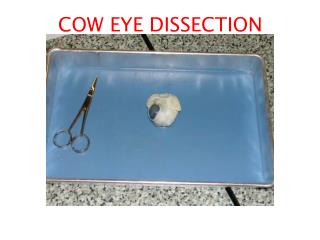
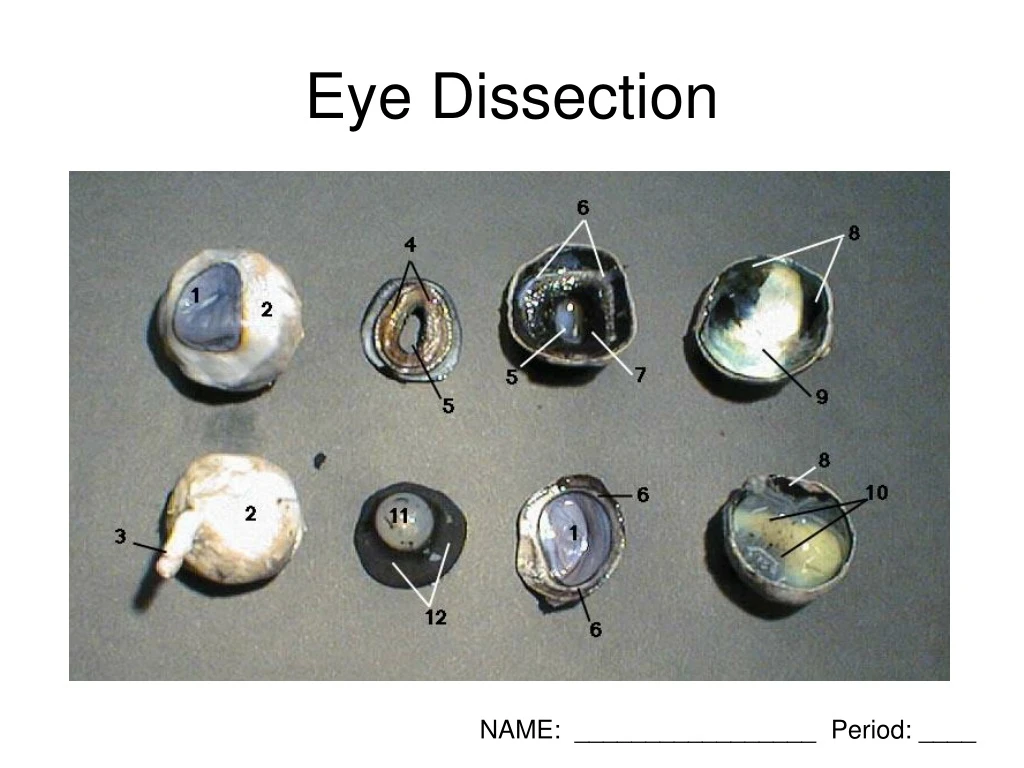
Eye Dissection NAME: _________________ Period: ____
The white part is the ________, the outer covering of the eyeball. The blue is the _______, which starts out clear but becomes cloudy after death.
Without moving your head, look up. Look down. Look all around. Six muscles attached to your eyeball move your eye so you can look in different directions Cows have only four muscles that control their eyes (superior and lateral recti). They can look up, down, left, and right, but they can’t roll their eyes like you can.
Now we are going to cut through the sclera and divide the eye in half, right around the middle. The cornea will be on the front half of the eye. The cornea is made of many layers of tissue.
If you look at your eye in a mirror, you’ll see a colored circle with a black spot in the middle. The colored circle is the ________. The black spot in the middle of the iris is the ___________, a hole through the iris that lets light into the eye. In dim light, the pupil __________, letting lots of light in. • The next step is to pull out the iris. The iris is between the cornea and the lens. It may be stuck to the cornea or it may have stayed with the back of the eye. Find the iris and pull it out. It should come out in one piece. You can see that there’s a hole in the center of the iris. That’s the pupil, the hole that lets light into the eye. The iris contracts or expands to change the size of the pupil. In dim light, the pupil opens wide to let light in. In bright light, the pupil shuts down to block light out.
Here is the back half of the eye. With the cornea and the iris out of the way, you can see the _______. It looks gray in this photo, but it’s really clear. The clear goo around the lens is the _________ _________. The eyeball stays round because it’s filled with this clear goo.
Here, the _______ works like a magnifying glass, making the words look bigger. The lens of the cow’s eye (like the lens of your eye) is shaped like the lens of a magnifying glass. It’s thicker in the middle than it is at the edges. This is called a _____________ lens.
Here’s the back of the eye with the lens and vitreous humor removed. It’s shaped like a bowl. On the inside of the bowl is a thin film with red blood vessels running through it. The _________ contains light-sensitive cells that detect light.
Here’s the inside of the back of the eye again. Behind the retina is a layer of shiny, blue-green membrane called the tapetum. This layer assists night vision by reflecting light back through the retina. You don’t have a tapetum, but cats and cows (and other animals) do. A cat’s eyes shine in the headlights of a car because of the tapetum.
Name the parts • ____________ 2. ____________ 3. _____________ 4. _______________ • ____________ 6. N/A 7. N/A 8. N/A • 9. _____________ 10. ____________ 11. ___________ 12. ____________