Download
1 / 27
290 likes | 534 Vues
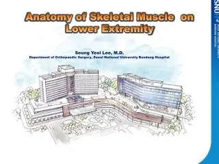
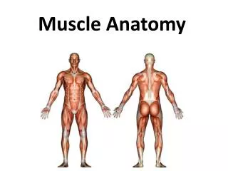
ANATOMY OF A MUSCLE. FROM FASCIA TO FILAMENT. Connective Tissue Coverings. Skeletal Muscle- Organ Composed of muscle, nervous, circulatory and connective tissue A Muscle is an organ with several levels of organization

E N D
ANATOMY OF A MUSCLE FROM FASCIA TO FILAMENT
Connective Tissue Coverings • Skeletal Muscle- Organ Composed of muscle, nervous, circulatory and connective tissue • A Muscle is an organ with several levels of organization • Each one is covered with its own type of connective tissue to hold its position
Levels of Connective Tissue • Aponeuroses- large sheets of connective tissue that attach to the facia of adjacent muscles • Facia- surround each entire muscle on the outside • Extends beyond the muscle to form tendons • Epimysium- right under facia • Perimysium- separates the muscle into smaller compartments called fascicle • Endomysium- separates each fascicle into individual muscle fibers
Levels of Organization • Muscular System V V • Muscle Group V V • Muscle V V • Fascicle (bundle of muscle fibers)
Levels of Organization • Muscle Fiber (from fascicle)- the muscle cell V V • Myofibril V V • Sarcomere (from one Z line to next Z line of myofibril) V V • Filaments (actin and myocin)
Skeletal Muscle Fibers • Muscle Fiber- a cell that responds to stimuli & relaxes when stimuli ends • Actually the fusion of many cells called a myoblast • Multinucleated due to this fusion • Other cellular structures have special names due to the fiber’s unique structure • Sarcolemma- outer covering • Sarcoplasm- inner fluid • Sarcoplasmic Reticulum- nework of internal channels • Transverse Tubules- channels that lead outside • Combination of two networks activate muscle contractions
Skeletal Muscle Fibers • Each fiber is very dense with mitochondria • Myofibrils- parallel threadlike subdivisions in each fiber • Fundamental in muscle contraction • Actin (thin) & Myosin (thick)- two types of protein filaments • The alternating of these filaments produces muscle striations • Muscle Striations- Two Main Parts • I Bands- light bands made up of thin actin filaments directly attached to structures called Z lines • A Bands- dark bands composed of thick myosin filaments overlapping with thin actin filaments • The H Zone is the middle of the A Band consisting of the M line (thickening of the myosin filaments) • Sarcomere- segment of the myofibril that extends from Z line to Z line
Figure 23.1: Sarcomere microanatomy: Relaxed (above) and contracted (below): • (a) actin (b) myosin • (c) Z-line (d) H-zone • (e) I-band (f) A-band • (green, diagonal arrows) H-band and I-band which change in length during contraction, (red, vertical arrows) A-band which does not change in length.
Motor unit • A motor unit is a single motor neuron and all of the corresponding muscle fibers it innervates. • When a motor unit is activated, all of its fibers contract. • Groups of motor units often work together to coordinate the contractions of a single muscle; • all of the motor units that subserve a single muscle are considered a motor unit pool.
ROLE OF ACTIN AND MYOSIN • Sliding Filament Theory- Muscle contractions result in actin and myosin filaments sliding across one another • Myosin molecule- two twisted protein strands • Cross bridges- globular heads running down its length • Myosin strand- Many myosin molecules • Actin molecule- globular structures that have binding sites for the myosin cross bridges • Actin strand- many actin molecules in a helix
Muscle Contracts When Myosin Crossbridges Attach to Actin and the Molecule Bends • The filaments slide together because myosin attaches to actin and pulls on it • Myosin head (H) attaches to actin filament (A), forming a crossbridge • After the crossbridge is formed the myosin head bends, pulling on the actin filaments and causing them to slide: • Muscle contraction is a little like climbing a rope. The crossbridge cycle is: grab -> pull -> release, repeated over and over
ATP is Required for Both Contraction and Relaxation of Muscle • ATP is the energy supply for contraction • It is required for the sliding of the filaments which is accomplished by a bending movement of the myosin heads • It is also required for the separation of actin and myosin which relaxes the muscle
STIMULUS FOR CONTRACTION • Action potential impulse sent from CNS causingCalciumIons to be pumped into the motor neuron • Calcium ionscauses the synaptic vesicles to open • Neurotransmitter acetlycholene (Ach) is released • Ach diffuses across the junction and binds to a receptor protien in the post synaptic membrane • This depolarizes the membrane, transfers the action potential to the muscle
Muscle Contraction and Relaxation • The action potential is received in the muscle membrane and travels through the transverse tubules to the sarcoplasmic reticulum (SR) • In response to the impulse Ca++ ions are released from the (SR) and goes to the actin filaments • This causes the myosin cross bridges to form linkages to the actin and a muscle contraction occurs • The contraction will continue as long as ATP is present and nerve impulses cause Ach release.
MUSCLES ARE NATURALLY IN A STATE OF RELAXATION • When the nerve impulse stops, Ach is broken down and sent back to the nerve for recycling • Also, Ach breakdown causes the Ca++ to go back into the SR and the actin-myosin linkages are broken, thus relaxation
Figure 23.1: Sarcomere microanatomy: Relaxed (above) and contracted (below): • (a) actin, • (b) myosin, • (c) Z-line, • (d) H-zone, • (e) I-band, • (f) A-band, • (green, diagonal arrows) H-band and I-band which change in length during contraction, (red, vertical arrows) A-band which does not change in length.
ENERGY FOR MUSCLE CONTRACTION • Muscle contractions take more ATP than is available in fibers • Creatine Phosphate- is able to make ATP from ADP and phosphate • Stored in the mitochondria OXYGEN SUPPLY & CELLULAR RESPIRATION • Oxygen for cellular respiration comes from red blood cells • Hemoglobin- pigment molecules in red blood cells that allows them to carry oxygen • Myoglobin- pigment in blood cells that also binds to oxygen OXYGEN DEBT & MUSCLE FATIGUE • Oxygen Debt- During activity, oxygen is used up and anaerobic respiration occurs • Anaerobic respiration results in the production of lactic acid • Muscle Fatigue- caused by a build up of lactic acid
MUSCULAR RESPONSES • Use single muscle fiber to observe • Threshold Stimulus- minimum strength of stimulus required to cause a muscle contraction • All-or-None Response- muscle fibers do not partially contract. Once the threshold stimulus is reached, a full contraction occurs. • Myogram- pattern a muscle contraction produces • Twitch- a single muscle contraction- 3 parts • Latent period- delay btn stimulus and response • Period of contraction- when muscle pulls • Period of relaxation- when muscle returns to original length • Summation- series of twitches that increase in strength before complete relaxation • Tectanic contraction- no relaxation between twitches
Recruitment of motor units- muscles are organized into motor units • Motor units are stimulated all-or none, one at a time until all the motor units for that motion are stimulated • Sustained contractions- when summation and recruitment are combined over time • Muscle tone- sustained contractions occurring continually in a resting muscle
SMOOTH MUSCLE • Contractile mechanisms for smooth and cardiac muscles are the same as those of skeletal muscles • Smooth muscle fibers- no striations • Multiunit smooth muscle- occur as separate fibers rather than in sheets • Irises of the eyes and blood vessels • Visceral smooth muscle- sheets of spindle shaped cells- more common • Walls of hollow organs- stomach, intestine, bladder • Peristalsis- rhythmic contraction of smooth muscles • Smooth muscle- slower to contract than skeletal muscle, but can maintain contraction longer with the same amount of ATP
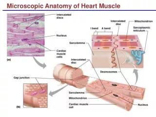
CARDIAC MUSCLE • Occurs only in the heart • Striated cells joined end to end • Long contractions • Intercolated Discs- cross bands that connect opposing ends of cardiac muscle cells • Cardiac muscle cells form a network and act as a single unit when the all-or-none stimuli response occurs • Cardiac muscle is self exciting and rhythmic
SKELETAL MUSCLE ACTIONS • Provide body movement • Origin & Insertion • Origin- immovable end of a muscle • Insertion- movable end of a muscle • Action- movement muscle causes • Dependent on attachments and type of joint
INTERACTIONS OF SKELETAL MUSCLES • Skeletal muscle function in groups • Prime Mover- muscle in group that provides most of the movement • Synergists- muscles that assist the prime mover • Antagonists- muscle that resist the prime mover and move in the opposite direction