Download
1 / 63
650 likes | 925 Vues
Anatomical Review of Muscle Cell anatomy. Fig. 12.1. Whole muscle = many muscle cells + CT. Fig. 12.15. Individual Muscle Cell—Anatomy Review. Fig. 12.6. Organization of actin and myosin filaments --Alternating and overlapping. Fig. 12.7. Organization of a sarcomere. Muscular System.

E N D
Fig. 12.1 Whole muscle = many muscle cells + CT
Fig. 12.15 Individual Muscle Cell—Anatomy Review
Fig. 12.6 Organization of actin and myosin filaments --Alternating and overlapping
Fig. 12.7 Organization of a sarcomere
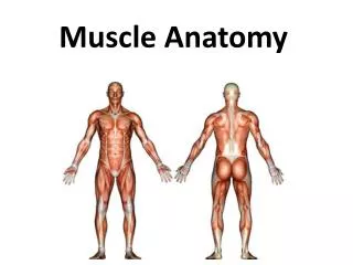
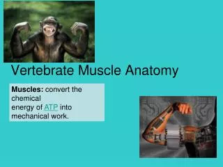
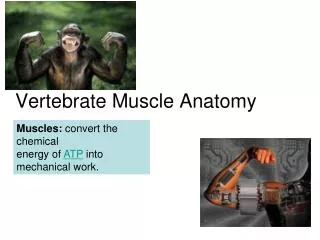
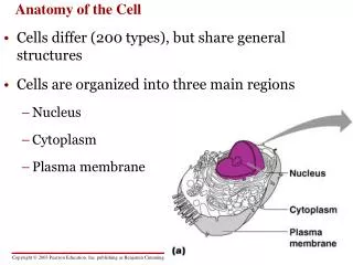
Muscular System • Skeletal Muscles and associated connective tissue • Skeletal muscle cells=muscle fibers FUNCTIONS • Produces movement • (through contraction of cells) • Important in verbal and non-verbal communication • Stabilizes joints and maintains posture • (through contraction of cells) • Produces body heat • (through high levels of cellular respiration)
OVERVIEW OF SKELETAL MUSCLE ACTIVITY BrainMotor Neurons, synaptic activity--ACh Action Potential & propagation Ca+ channel activity ATP production/consumption sliding of actin & myosin filaments production of force and movement (sometimes)
Voluntary Motor Activity Originates in Frontal Lobe of cerebral cortex
Voluntary Muscle Contraction: • Neuron Activity Begins Frontal Lobe: • Upper motor neuron • Decussates in medulla (~80%) • Travels down spinal cord through anterior or lateral corticospinal tracts to lower motor neuron • Synapse with lower motor neuron • Lower motor neuron travels through nerve to effector muscle • Forms synapse—Neuromuscular Junction—with muscle • NT= Ach binds to nicotinic receptors
Neurological Control of Skeletal Muscle • CNS (brain and spinal cord): generate motor commands that will signal muscle cells to contract • Voluntary Activity • Frontal lobe: initiates voluntary muscle activity • Basal nuclei: coordinates voluntary muscle activity • Thalamus: involved with coordination of voluntary muscle activity • substantianigra: coordinates muscle activity (inhibits antagonistic muscles) • Cerebellum: coordinates muscle activity (makes adjustments based on current body position) • Cranial reflexes • Generate involuntary, reflexive muscle use to specific stimuli. Integrating center is in brain • Spinal Cord • Spinal reflexes • Generate involuntary, reflexive muscle use to specific stimuli. Integrating center is in spinal cord • Lower motor neurons (PNS) directly innervate muscle cells • CNS initiated commands are relayed (through synapses) to lower motor neurons which carry A.P.s from CNS to the individual muscle cells they innervate.
Neurological Control of Skeletal Muscle • All Skeletal Muscle cells are directly innervated by a motor neuron • Neuromuscular Junction: • The chemical synapse between a motor neuron and a muscle fiber (cell) • Chemical synapse, always excitatory • Motor Units: a motor neuron and all the muscle cells it innervates • Multiple fibers innervated by same neuron • They contract together as a unit
Fig. 12.4 This neuron is also a lower motor neurons
Key NMJ concepts Chemical synapses (as described in neuron physiology unit) Neurotransmitter: ACh Receptor: nicotinic Receptor Action: ACh opens ligand gated Na+ channel, Na+ enters cells depolarizing itend plate potential Short-lived due to action of ACh’ase NMJ are always excitatory A single AP almost always releases enough ACh to bring the motor end plate/muscle cell to threshold
Another representation of the events at a neuromuscular junction
EXCITATION-CONTRACTION COUPLING: from AP formation at synapse to actin-myosin interaction • AP propagates across PM (sarcolemma of muscle cell) • VG Na+ channels • Just like an AP in axon • AP travels down T-tubels • VG Ca+ channels (aka DHP receptors) open • DHP receptors are coupled/linked to Ca+ release channels • Ca+ release channels (aka ryanodine receptors) open • Ca+ floods into cytoplasm • Ca+ binds actin filament allowing actin-myosin interaction
Fig. 12.16 Visual representation of excitation-contraction coupling
Fig. 12.17 Flow Chart of excitation-contraction coupling events
Fig. 12.13 ACTIN FILAMENTS Covers up binding sites for myosin heads, can move to expose binding sites Ca+ binds Has binding sites of myosin head, will be bound by myosin during interaction/contraction
Fig. 12.10 MYOSIN FILAMENT STRUCTURE • Myosin Head: • Binds Actin • Have binding site for ATP • Will grab, pull on, and detach from actin
Fig. 12.9 • Filament Interaction: • Myosin grabs and pulls on actinfilaments slide across one another • Zone of filament overlap increases • Sarcomeres get shorter cell shortens=contraction
FILAMENT INTERACTION: SLIDING FILAMENT THEORY OF MUSCLE CONTRACTION • Myosin and actin filaments interact • Myosin pulls on actin • Filaments slide past one another increasing zone of overlap • Sarcomeres get smaller • Results in contraction of muscle and production of tension (i.e., pulling force)
When Ca+ binds troponin, tropomyosin moves to expose myosin binding sites as shown in diagram NOTE: This is show as if you were viewing the filaments along their short axis—different perspective then other diagram
Production and Control of Tension • Contraction produces pulling force known as tension
Twitch: a single contraction. The result of a single AP/excitation contraction coupling event • Latent: • AP propagation, Ca release, Ca build up in sarcoplasm • Contraction: • Active cross bridging/contraction, Ca+ available • Relaxation: • Ca+ decreasing in sarcoplasm, diminished and eventual lack of crossbridging/contraction
Figure 9.41 Relationship between time of stimulus, AP, and tension
Factors that influence tension (i.e., strength of contraction) • Action Potential (stimulus) frequency • Number of active fibers/number of motor units activated • Fiber length (amount of actin-myosin overlap)
Figure 9.19 Green: single twitches, complete relaxation between Orange: partial relaxation between two stimuli stimuli wave summation—second contraction stronger than first Purple: two stimuli with no relaxation between contraction stronger than that with a single stimulus
Summation (temporal/frequency) • Increased stimulation rate increased tension/strength • Build up/availability of Ca+ in cytoplasm • Incomplete/unfused tetanus—stimulation frequency allows partial contractions • Complete/fused tetanus – stimulation frequency does not allow any relaxation phase.
Recruitment = strength of contraction proportional to number of motor units activated • E.g., ↑ motor unit = ↑ tension/strength • Rotating through motor units allows prolonged contraction with reduced fatigue
Optimal resting length = optimal overlap of filaments ↑ cross bridging ↑ tension • Too long = too little overlap not enough crossbridging ↓ tension • Too short = no room left to contract & fiber mis-alignment ↓ tension ˃˂↑↓
Energetics ATP needed for: • Energizing head • Detaching head from myosin • Power Ca+ pumps that transport Ca+ into SR (from cytoplasm) ATP production through: • Aerobic respiration • Anaerobic respiration • Creatine Phosphate
Stored energy sources and how much muscle contraction they can sustain.
Resting Muscle • Primary substrate plasm fatty acids • ATP production ˃ ATP consumption/demand • Surplus ATP used to: • Creatine CP • Glucose glycogen ˃˂↑↓
Moderate activity • Substrates = plasm fatty acids & glucose/glycogen • ATP production can meet ATP consumption/demand • Aerobic respiration dominates