Download
1 / 34
360 likes | 705 Vues
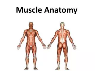
Anatomy of the Muscle. Mr. Neuberger. Types of Muscle. Skeletal Muscle Defined as voluntary, striated muscle that’s attached to one or more bones Striations are overlapping arrangement of the contractile proteins Muscle cells are called fibers or myofibers due to their length Smooth Muscle

E N D
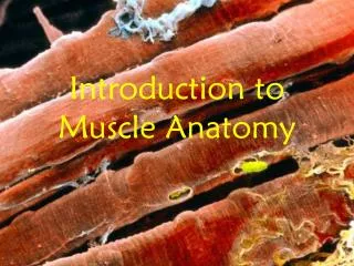
Anatomy of the Muscle Mr. Neuberger
Types of Muscle • Skeletal Muscle • Defined as voluntary, striated muscle that’s attached to one or more bones • Striations are overlapping arrangement of the contractile proteins • Muscle cells are called fibers or myofibers due to their length • Smooth Muscle • Cardiac Muscle
Functions of Muscle • Movement • Allow us to move from place to place and move individual body parts • Also aid in the movement of body contents during breathing, circulation, digestion, etc • Stability • Help maintain posture, prevent unwanted movement • Antigravity muscles • Stabilize joints by constant tension on tendons and bones • Heat Production • Muscles are responsible for as much as 85% of the body’s heat production, vital to function of all enzymes and therefore to all metabolism • Control of Body Openings and Passages • Eye • Mouth • Urethra and Anus
Properties of Muscle • Excitability • Responsiveness • Property of all living cells, developed to the highest degree in muscle and nerve cells • Exhibit electrical and mechanical responses to stimuli • Conductivity • Local electrical excitation produced at the point o muscle stimulation is conducted throughout entire plasma membrane • Contractility • Shorten substantually when they are stimulated • Pull on bones(skeletal muscles) as a result • Extensibility • Can stretch as much as three times their contracted length • Most cells in body show little to no extensibilty • Elasticity • When tension in muscle fiber is released, it recoils to its original length
Connective Tissue • Endomysium • Thin sleeve of loose connective tissue that surrounds each muscle fiber • Creates room for blood capillaries and nerve fibers • Perimysium • Connective tissue sheath that wraps muscle fibers together in bundles called fascicles. Carries the larger nerves and blood vessels as well as stretch receptors called muscle spindles • Epimysium • Fibrous sheath that surrounds entire muscle • Grades into the fascia • Fascia • Sheet of connective tissue that separates neighboring muscle or muscle groups from each other and subcutaneous tissue
Muscle Shapes • Fusiform • Thick in the middle and tapered at the end • Biceps brachii is an example • Parallel • Fairly uniform width and parallel fascicles • Rectus abdominus • Span longer distances and shorten more than other muscles but produce limited force • Triangular(Convergent) • Fan-shaped, broad at origin and narrow at insertion • Relatively strong due to the number of fibers in the wide part of the muscle • Pectoralis major is an example
Muscle Shapes cont. • Pennate • Feather-shaped • Fascicles insert obliquely on a tendon that runs the length of the muscle • Unipennate- approach tendon from one side • Semimembranosus • Bipennate- approach tendon from both sides • Rectus femorus • Multipennate- multiple “feathers” with quills approaching one point • Deltoid • Circular • Form rings around body openings • Orbicularis oculi
Muscle Attachments • Indirect • The muscle ends short of the bony destination • The gap is bridged by a fibrous band called a tendon • Strong continuity from muscle to bone • Aponeurosis- tendon is broad sheet, example is in the palm of the hand • Direct(Fleshy) • To the naked eye, appears as if muscle is attaching directly to bone • Small gaps filled by collagen fibers • Some muscle will insert on fascia or tendon of another muscle, not bone
Functional Groups of Muscles • Prime Mover(Agonist) • Muscle that produces the most force during a joint action • Synergist • Aids the prime mover • Stabilizer • Antagonist • Opposes action of prime mover • Antagonistic pair (triceps and brachialis) • Fixator • Prevents the bone from moving • Scapula during a dumbell curl
Intrinsic vs Extrinsic • Intrinsic muscles are entirely contained within a particular region • Extrinsic muscles act upon a designated region but has its origin elsewhere
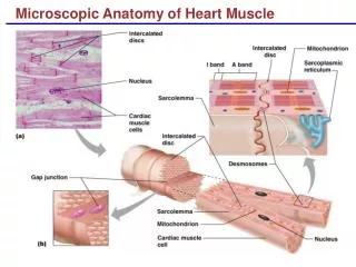
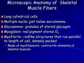
Ultrastructure of Muscle Fibers • Sarcolemma • Plasma membrane of the muscle fiber • Sarcoplasm • Cytoplasm of the muscle fiber • Myofibrils • Sarcoplasm is occupied by long protein bundles called myofibrils • Myoglobin • Red pigment within the cells that stores oxygen needed for muscular activity • Sarcoplasmic Reticulum • Smooth ER, forms a network around each myofibril
Ultrastructure of Muscle Fibers cont. • Terminal cisternae • Dilated end-sacs cross the muscle fiber from one side to the other • Transverse (T) Tubules • Sarcolemma turns inward at many points to form tunnels called T tubules, penetrate through the cell and emerge on the other side
Myofilaments • Thick Filaments • Made of several hundred molecules called myosin • One half moves right, one half moves left • Thin Filaments • Combination of two intertwined strands of protein • Tropomyosin counteracts action of myosin by blocking it • Elastic Filaments • Made of titin • Flank each thick filament and anchor it to the Z disc
Contractile Proteins • The two main proteins are Myosin and Actin • They do the work of shortening the muscle fiber • Tropomyosin and Troponin are called the regulatory proteins • Act like a switch to determine when it can contract and when it cannot
Striations and Sarcomeres • A bands • Anisotropic • Consists of thick filaments lying side by side and partially overlap the thin filaments • I bands • Isotropic • Consists of thin filaments only, lying side by side • H band • Lighter region of the A band where thin filaments do not reach
Striations and Sarcomeres cont. • M line • Line where the thick filaments originate • Z disc(line) • Bisects the I band • Dark, narrow, and provides anchorage for the thin filaments and elastic filaments • Sarcomere • Segment of the myofibril from one Z disc to the next
The Nerve-Muscle Relationship • Skeletal muscles are innervated by somatic motor neurons • Cell body lies in the brain stem and spinal cord and their axons, somatic motor fibers, lead to the skeletal muscles • Neuromuscular Junction • The nerve fiber branches and establishes several points of contact • Point where the nerve and muscle fiber meet, there is an indentation • Synaptic Knob- bulbous swelling at each synapse • Space between is called synaptic cleft
The Synaptic Knob • Contains spheroid organelles called synaptic vesicles • Filled with a chemical called acetylcholine (ACh) • When a nerve signal arrives at the synaptic knob, some of the vesicles release their ACh by exocytosis • Diffuses across the cleft and binds to membranes called ACh receptors on the sarcolemma • Sarcolemma has infoldings called junctional folds that increase the membrane surface area
Basal Lamina • Thin layer composed partly of collagen and glycoproteins, which separates it from the surrounding connective tissue • Passes through the synaptic cleft and virtually fills it, also covers the Schwann cell of the NMJ • Acetylcholinesterase is found in both the sarcolemma and basal lamina • Breaks down the ACh after it has stimulated the muscle cell, this is important for turning off the muscle contraction
The Motor Unit • The muscle fibers innervated by a singular nerve fiber all function in unison and they are called a Motor Unit • The muscle fibers of a single motor unit are not all clustered together but they are dispersed throughout a muscle • When they are stimulated, they cause a weak contraction over a wide area, not just localized to a twitch • Roughly 200 muscle fibers per motor neuron • Small motor units have 3-6 muscle fibers per nerve fiber • Large motor units have roughly 1000 muscle fibers per nerve fiber
The Blood Supply • The muscular system, as a whole, receives about 1.25L of blood per minute at rest (1/4 of the blood pumped by the heart) • Heavy exercise will cause the muscles to receive 11.6L/min (3/4 heart output) • Working muscle has a great demand for glucose and oxygen • Blood capillaries will run through the endomysium to reach every muscle fiber • These capillaries will coil when the muscle is contracted so that they have enough slack for when the muscle stretches back out
Contraction and Relaxation • 4 stages, 10 steps • Excitation • Excitation-Contraction Coupling • Contraction • Relaxation
Excitation • (1) A nerve signal arrives at the synaptic cleft • (2) Synaptic vesicles release ACh , which diffuses across the synaptic cleft and binds to receptors on the sarcolemma • Opens ion gates in the sarcolemma, resulting in sodium and potassium movements through membrane that electrically excites the muscle fiber • (3) A self-propagating wave of excitation spreads down the length of the fiber and into the T tubules
Excitation-Contraction Coupling • (4) Electrical events in the T tubules lead to the opening of calcium gates in the sarcoplasmic reticulum • Releases calcium into the cytosol • (5) Ca²+ attaches to the troponin molecules in the thin filaments. Troponin induces the tropomyosin to shift position to fall into the groove between the two F actin filaments and exposing the active sites on the G actin • (6) Myosin has been “waiting” in a flexed position with a molecule of ATP bound to its head. • ATP + myosin ATPase = ADP + P
Contraction • (7) Myosin forms a link, cross-bridge, with one of the active sites of actin, and releases ADP and P • (8) Myosin flexes and tugs on the thin filament. The thin filament slides a short distance along the thick filament, called the power stroke. • Myosin binds to a new ATP, lets go of the thin filament, breaks down the ATP, and recocks. • As long as the muscle is contracting, the thin filament is never entirely released, other myosin heads hold on • Steps 6-8 will occur repeatedly and produce the sliding filament theory • Can shorten up to 40%
Relaxation • (9) Stimulation of the muscle must stop in order for the muscle to relax. • The motor neuron stops firing and releasing ACh, so the muscle fiber is no longer electrically excited • (10) The sarcoplasmic reticulum reabsorbs the calcium ions and stores them until the next time the muscle is stimulated • Without calcium, troponin moves back into the position that blocks the active sites and prevents myosin-actin cross-bridges from closing
Muscle Growth • Skeletal muscle fibers are incapable of mitosis. • Exercise stimulates the muscle to produce more myofilaments, thus myofibrils grow thicker • At a certain point a large myofibril splits longitudinally • Well-condition muscles also develop more mitochondria, myoglobin and glycogen, and a greater density of blood capillaries
Slow twitch (Type I) Fibers • Abundant mitochondria, myoglobin, and glycogen • Well adapted to aerobic respiration • Means for making ATP but does not generate lactic acid as byproduct • Take longer to contract, but do not fatigue easily
Fast Twitch (Type II) Fibers • Rich in enzymes for anaerobic fermentation • Process that is independent of oxygen but produces lactic acid • React to a stimulus faster but fatigue easily • Quick responses, not endurance