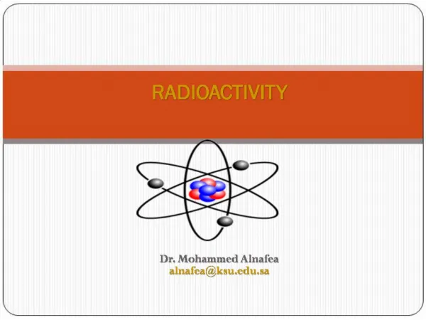
Chapter 2 Radioactivity
Chapter 2 Radioactivity. The Atomic structure. RADIOACTIVITY. Objectives: Introduction Four types of radioactive decay Penetration power of radiation Natural Decay Law Radiation units Biological effects of radiation Pocket dosimeter Medical applications

Chapter 2 Radioactivity
E N D
Presentation Transcript
RADIOACTIVITY Objectives: Introduction Four types of radioactive decay Penetration power of radiation Natural Decay Law Radiation units Biological effects of radiation Pocket dosimeter Medical applications References:1-Medical Physics textbook by Cameron
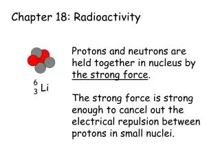
What is Radioactivity? - It is the property of an atom to change into atoms of another element. - Spontaneous change is called decay. - Radioactivity is a property of the material and can be either natural or artificially induced. - Radiation in simple terms is the “Transfer of energy through space and matter”. Unstable elements are called radionuclides or radioisotopes which are characterised by: 1- Half-life (T½) [Time needed for half of the radioactive nuclei to decay] 2- Mode(s) of decay [α, β, γand spontaneous fission] 3- Type of radiation emitted 4- Products of the decay which may also be radioactive. Radioactivity is a naturaland spontaneous (decay) process by which the unstable atoms of an element emit or radiate excess energy in the form of particlesand waves. These emissions are collectively called “ionizing radiations”. Depending on how the nucleus loses this energy either a lower energy atomof the same form will result, or a completely differentnucleus and atom can be formed.
Radiology is concerned with studying the effect of radiation to the human body for diagnostically and therapeutically application. In order to evaluate the advantages and disadvantages of the various medical applications of radiation and its limitations, this requires an understanding of the following concepts: 1- The basic nature of radiation. 2- Interaction between radiation and matter. 3- Radiation detection. 4- Biological effects of radiation. Nuclear energy is released by the splitting (fission) or merging (combining) together (fusion) of the nucleiof the atom (s). Nuclear energy is released via three exothermic processes: 1- Radioactive decay: A neutron or proton in the radioactive nucleus decays spontaneously by emitting either particles, electromagnetic radiation (gamma rays), neutrinos or all of them. 2- Fusion: Two atomic nuclei fuse together to form a heavier nucleus. 3- Fission: The breaking of a heavy nucleus into two (or more rarely three) lighter nuclei.
Radioactivity is associated with the transmutation of the nucleus from one element to another. Thus, for example, when radium (Ra) emits an alpha (α) particle, the nucleus is transformed into radon (Rn). These emissions are collectively called ionizing radiations. Depending on how the nucleus loses this energy either a lower energy atom of the same form will result, or a completely different nucleus and atom can be formed. What is Ionizing Radiation? - It is a radiation that has the ability to strip electrons from atoms, this process is calledionization. - Ionization can initiate biological damage so slight as to be totally unnoticeable or so severe as to cause death. ينزع
Main ionizing radiation are: , , , x-rays and neutrons [neutron p++ - (proton & beta particle {negatron}), neutrons don’t ionize directly, their energy range from 10-2 to 107 eV]. Note: eV is the unit used to measure the energy of a charged particle, and it is defined as “The energy an electron can gain when it is accelerated through a potential difference of 1 (V)”. What is Non-ionizing Radiation? Does not have enough energy to remove (strip) electrons from atoms, although it can interact with matter through other processes, some of which may be detrimental Sunburn is a good example of the effect of non-ionizing radiation. Sun is composed of 74% by mass hydrogen (11H0), an element whose nucleus is a single proton; energy is released when 4 protons combine into a helium (42He2) nucleus, a process in which two of them are also converted to neutrons (10n1). .(ضار)
Types of Atoms Unstable Stable Metastable 9943Tc56 Technetium Protons = 43 Neutrons= 56 Mass number = 99 235 U 92 4 He 2 A=Mass number Z= Atomic number 143 2 Uranium Protons = 92 Neutrons = 143 Mass number =235 Helium Protons = 2 Neutrons = 2 Mass number = 4 • - For nucleus to be stable a certain number of neutrons is required if this number is increased or decreased than required, then the nucleus will become unstable and start to disintegrate (decay) to reach a stable state. • Atoms with completely filled orbits [atoms of the so-called noble gases: helium, neon, argon, krypton, and xenon(133Xe)]cannot share electrons with other elements, therefore they are chemically most inert. • - Some man-made radionuclide emit types of radiation not emitted by natural radioactive substance such as technetium (Tc), which means that this element is half-stable. • - A metastable radionuclide decay by emitting γ rays only, and its daughter nucleus differs from its parent in having less energy.
The nucleus of mass is characterized by atomic number Z protons of mass mp and N neutrons of mass mn. The mass number A = [Z+N]. Protons and neutrons are closely related and are sometimes collectively known as nucleons. A simple description of nuclear binding energyis the energy required to break apart, split, or break down, the nucleus of the atom into its component parts (nucleons). Nuclear binding energy (binding energy of nucleons into a nuclide) can be easily computed from the easily measurable difference in mass of a nucleus, and the sum of the masses of the number of free neutrons and protons that make up the nucleus. Once this mass difference, called the mass defect or mass deficiency, is known, Einstein's mass-energy equivalence formula can be used to compute the nuclear binding energy of any nucleus: E =Δmc² Proton Nucleus Neutron Δm = mass defect, and c = speed of light in a vacuum. From this formula, adding energy increases mass, removing energy, decreases it. Δm=(sum of masses of protons and neutrons)-(measured mass of nucleus)
The difference between the actual mass of the nucleus (mnuclide) measured in amu and the number of nucleons (A=Z+N) is called mass excess. Mass excess = mnuclide– A The mass of the atom's nucleus (mnuclide) is always less than the sum of the individual masses of the constituent protons and neutrons (A). This notable difference is a measure of the nuclear binding energy, which is a result of forces that hold the nucleus together. A general and simple description of nuclear binding energyis the energy required to break apart, split, or break down, the nucleus of the atom into its component parts (nucleons). If the binding energy for the products is higher when light nuclei fuse, or when heavy nuclei split (break down), either of these processes will result in a release of the "extra" binding energy, and this energy is referred to as nuclear energy. It is also loosely called nuclear power.
8.8 58F 26 26 Iron is the third most tightly bound elements, its binding energy/nucleons ≈ 8.8 MeV 4 62 Ni 28 Yield from nuclear fission Elements heavier than Fe can yield energy by nuclear fission Yield from nuclear fusion 118 235 The main and highest peak in the curve is at iron (most tightly bound element) then the curve will slowly decreases, also a narrow isolated peak at helium (4He), is very stable. The heaviest nuclei in nature is uranium238U , which is unstable, having a lifetime of 4.5 billion years, close to the age of the Earth. Uranium is still relatively abundant; it may be formed in an explosion, preceding the formation of the solar system. In each of these, radioactive decay produces "daughter isotopes" which are also unstable, starting a chain of decays which ends in some stable isotope. Nickel (62Ni) isotope is the most tightly bound of nuclides.
Mass is often expressed in term of amu (atomic mass units) which is defined as 1/12 of the mass of carbon atom [126C6] by the following equation: 1/12 x12/No=1/(6.022 x 1023)=1.655 x 10⁻²⁴ (g) ≈1.66 x 10⁻²⁷(Kg)=931.25 (MeV) [No = Avogadro’s number = 6.022 x10²³ (g-1) = 6.022x1026 (Kg-1)] As higher the binding energy (BE) is as more stable the nucleus is. To calculate the "binding energy" we use this formula: BE= P(mp + me) + N.mn - mnuclide where P isthe number of protons of the radionuclides and N its number of neutrons. We take mp = 938.2723 MeV, me = 0.5110 MeV, mn = 939.5656 MeV and mnuclideis the actual mass of the nucleus in MeV.
The isotopes of a given element are chemically identical [i.e., they participate in the same chemical reactions but they could be distinguished from each other because their mass number (A) are different. Nuclei with equal number of neutrons and different A, Z are called isotones 177N10, 188O10, and 199F10 Nuclei with equal mass number but different Z, N are called isobars 187N11, 188O10 and 189F9 All these nuclei have different binding energies because they differ in the number of protons and neutrons from each other.
Four types of radioactive decay: 1) Alpha (a) decay [its charge is +2] - 24He (Helium) nucleus (2p + 2n) ejected. The binding energy of helium is appreciable, and seems to be the energy source of the Sun and of most stars. 2) Beta () decay [charge -1]- change of nucleus charge, conserves mass [their are three types of beta decay: -, + and electron capture (EC)]. 3) Gamma (g) decay [charge is zero] - photon emission, no change in A or Z 4) Spontaneous fission which generates two smaller nuclei. Unstable nuclei undergo transformation by the emission of energetic radiation such as: (1) alpha (α)particles,which are high-speed helium nuclei consisting of two protons and two neutrons; (2) beta (β)particles, which are very high-speed electrons; and (3) gamma (γ) rays, which are highly energetic photons.
The nucleus can be in higher excitation if it rotates (rotational energy), if it vibrates (vibrational energy) or if the single particles are in higher quantum mechanically allowed states (single particle excitation). -emission occurs by de-excitation of a high excitation level of the nucleus to the ground state. The energy difference between the two excited states corresponds to the energy of the -radiation. (High excitation level) Cobalt =2.823-0.318 =2.505-1.173 = 1.332-1.332 Nickel Nickel is the most tightly bound nucleus (per nucleon), followed by (5826Fe32) and (5626Fe30).
A is mass number = (Z + N) Z is Atomic number= protons number : decay is of three types 1) - decay [Blue ball indicate neutron and red ball indicate protons] electron antineutrino - A Z 2 1 n P+ Negatron P+ P+ - Neutron rich atom is required. - Converts one neutron into a proton and emit electron, negative beta particle (e-= β-). This is because neutrons are more massive than protons. - No change of A, but different element is formed. - Release of electron antineutrino. n n Hydrogen Helium 2) + decay Positron neutrino 5 6 - Proton rich atom is required. - Converts one proton into a neutron and emit positron, positive beta particle (e+ = β+)[for energetic reason and not to free particles]. -The conversion of protons to neutrons is the result of a weak (nuclear) force. - No change of A, but different element is formed. - Release of electron neutrino (ve). Carbon Boron
3) Electron capture (EC) K-electron n n p p p p p n n n n n p p Lithium Beryllium Parent Daughter EC 73Li + ve Proton rich nucleus 74Be + e- 4 3 (neutrino) If the mass difference between parent and daughter is less than 1.022 MeV (the mass of 2 electrons) a proton-rich nucleus may still convert protons to neutrons by the process of electron capture, in which a proton simply captures one of the atom's K orbital electrons (K electron), emits a neutrino, and becomes a neutron (i.e., number of protons decrease while, number of neutrons increase).
In -decay a neutron is converted into a proton by electron emission (-decay) and electron antineutrino (ve ), with no change in mass number (A) as shown in the example. (β-) (negatron) Radium Actinium A 140 139 Z (β-) Neutron rich atom
Most positrons emitters are produced by cyclotron, which accelerate positive particles to bombard the target material. To emit positron a proton is converted into a neutron by positron (antiparticle of the electron) emission (+decay) with no change in mass number. This is permitted if enough energy is available between parent and daughter nuclides to do this (the required energy difference is equal to 1.022 MeV, which is the mass of 2 electrons). (β+) Protactinium (Parent) Thorium (Daughter) 139 140 (Antiparticle of the electron) (β+) + Proton rich atom Cyclotron =محطم الذره β - particles have a continuous spectrum of energies because they share their energy with the neutrino.
In the -decay processes the nucleus reduces its mass by emitting a 42He2 (helium) nucleus (-particle) to reach a less massive state. -decay occurs in heavy massive nuclei. The kinetic energy of the emitted α-particles is determined by the mass of the parent (Ra) and daughter (Rn) systemwhich is a member of the uranium series. Parent - 138 (Radium) The released energy is translated into the kinetic energy of the emitted α-particle and the heavy recoil nucleus. α α Radium Radon Radon is an important terrestrial source of ionizing radiation because, although on average it is very rare. Radon is a decay product of uranium, which is relatively common in the earth’s crust. γ Daughter 136 Radon 138
Nucleus reduces its mass by emitting 42He2(α particle) Spontaneous (α) of Plutonium nucleus Plutonium Uranium 144 146 p n n p 2
Penetration power of radiation Note: that neutrons is the most penetrating as their energy range from 10-2 to 107 eV
Rang of Alpha and Beta radiation in the medium GM counter Medium Rangeof radiation particles (R) is defined as “the maximum distance traveled in the absorber material by the most energetic radiation particles”.
Interaction of radiation with matter Gamma (γ) Alpha (α) Beta (β) 1- Photoelectric effect 2- Compton scattering 3- Pair production Since the alpha particle is basically a helium nucleus (24He) it is the largest and most massive type of radiation. The major energy loss mechanism for alpha particles is electronic excitation and ionization. The specific ionization of an alpha particle is very high, in the order of thousands of ion pairs per centimeter of air. Beta particles are charged particles with relatively light mass. There are 2 main mechanisms of interaction: 1- Excitation, Ionization 2- Bremsstrahlung
Natural Decay Law The rate of the decay process is determined by the activity A (number of decay processes per second) of the radioactive sample. [A= Disintegration/second= dps] The activity [A(t)] is proportional to the number of radioactive nuclei (radionuclide), t is the time. is the decay constant = ln2/t = 0.693/t A(t) = Final activity Initial activity= A(t0)
Each radionuclide decays at a fixed rate commonly indicated by the half-life (t1/2). Each radioisotope has a unique and unchangeable half-life. Initial activity λ = ln2/t = 0.693/t -λt N = No e Number of nuclei N, the number of nuclei present at time t. Nothe initial number of nuclei. Activity of sample versus time plotted on a linear scale Activity of sample has an exponential relation as: A = λN= Aoe-λt Ais the activity (disintegration/second), Ao is the initial activity, λ (hour-1) is the decay constant and t is the time in hour since the activity was Ao. The unit of activity (disintegration per second) was named Becquerel (Bq), another unit used is Curie (Ci).
Radioactivity versus time for the common radionuclide on a semi-log scale 100 50 25 12.5 Radioactivity % 6 A = Aoe-λt 1 0 6 12 18 24 Time (hr) The straight line on the semi-log graph indicate that the decay is exponential as shown in the equation. If the graph shows a straight line then only one radionuclide is present. While, a curved line indicate the presence of more than one radionuclide.
N(to), A(to) are the initial number of radionuclide and initial activity, respectively. Half life (t1/2) of a radionuclide: Is the time required for the radioactive material to decrease by one - half of their initial value. t1/2= ln2/λ = 0.693/λ Number of nuclei present at half life time = N(t½) There is an inverse correlation between half life time (t1/2) and decay constant (λ) for each radionuclide. Dividing both sides by N(t0) [Take ln of both sides] [Since λ = ln2/t1/2 then 1/λ = t1/2/ln2] ln [e-λt1/2] = ln1/2= -λt1/2 = t1/2 The life time () of a nucleus is defined by: 0.693
Radiation units Activity Units: The unit for measuring the activity of radiation source [number of decays (disintegration) per time] is Becquerel (Bq) or Curie (Ci): 1 Bq = 1 decay/ second = 2.7 x 10⁻¹¹ (Ci) One curie is defined as: The activity of 1 gram of pure Radium 1 Ci = 3.7 x 10¹⁰ (decay/s) = 3.7 x 10¹⁰ (Bq) Dosimetry Units: the measurement or calculation of energy deposition to material by radiation”. Due to the interaction between radiation and material ionization occurs in the radiated material (Energy transfer from the high energetic radiation photons or particles to atomic electrons in the material).
Unit for exposure (E) is the Roentgen [R] which is defined by the “ionization between EM-radiation and air”. Roentgen is defined as” the amount of EM-radiation which produces in one gram of air 2.58 x10⁻⁷ (Coulomb) at normal temperature (22°C) and pressure (760 mmHg) conditions” R = 2.58 x 10⁻⁷ (C/g) = 2.58 x 10⁻⁴ (C/Kg) • Roentgen is used to measure the amount of energy in a photon beam just before entering the skin of a patient. It is defined only for measurement in air. And it applies only to x and gamma rays up to energies of about 3 MeV. Exposure rate (ER)Is the radiation intensity measured in [Roentgen/hour] it can be related to the activityAof a source (in units mCi) via this equation: ER = (Γ) x A (R/h) i.e., ER is directly proportional to activity [A in (mCi)] d2 Гis the exposure constant for a particular radionuclide in [(Rcm2)/(hmCi)], and d is the distance between source and material in units (cm). The exposure constant is characteristic for the different radiation sources.
Absorbed dose (D) of radiation in any kind of material depends on the typical ionization energy of the particular material. The absorbed dose is defined as “the energy deposited in any medium (tissue) due to radiation absorption”. It is measured in ergs/gram or joules/kg = Gray = Gy = 100 rad Absorbed dose therefore, clearly depends on the energy loss behavior of various kinds of radiation: D = E x W1P Where, E is energy deposited in any medium, W1P is the average ionization energy for air, W1P 34 (Joul/Coulomb). Example:Calculate the absorbed dose in rad for 1(R) exposure of (EM) radiation, knowing that 1 R = 2.58 x 10⁻⁴ (Coulomb/kg). D = E x W1P =1R x 34 (J/C) = 2.58 x 10⁻⁴ (C/kg) x 34 (J/C) = 87.72 x 10⁻⁴ (J/kg) = 87.72 x 10⁻⁴ (Gy) = 87.72 x 10-4 x 102 rad = 0.8772 rad
Absorbed dose rate (DR) If radiation of energy Er and an activity A is completely absorbed in material of mass (M) Kg. The absorbed dose rate DR is: DR = A (Er / M) (Bq.MeV/Kg) Example: A patient receives an injection of 1.1x 10⁸ (Bq) of 131I, which accumulate in the thyroid gland (Mthyroid = 20 g). The mean energy of the emitted radiation is Er = 300 KeV. What is the dose rate to the thyroid? Note: MeV =106 (eV) =1000 KeV hence, (KeV) = 1/1000 (MeV) Answer: DR = A (Er / M) M thyroid = 20 (g) = 0.02 (Kg), Er = 300 KeV = (300/1000)= 0.3 MeV (Absorbed dose rate) = DR = 1.1 x 108 x (0.3/0.02)= 16.5 x 10⁸ (Bq.MeV/Kg)
Equivalent dose (H): Is the absorbed dose in tissue or organ of T weight for the type and quality of radiation R. H is calculated by multiplying absorbed dose (DT,R) averaged over organ or tissue due to radiation by the appropriate radiation weighting factor (WR). • H = DT, R x WR (Sievert =100 rem ) • If DT,R is in rad, the dose equivalent (H) is in rem and If DT,R is in Gy then H will be in Sv. • Every radiation type has its own weighting factor WR • Effective dose (Hε): Accounts for the effectiveness or quality of the radiation. It also includes a factor representing the sensitivity of the tissue to the radiation, so it also accounts for how the tissue might react. H is defined as “The sum of the products of the equivalent doses (H) to the organ and tissues exposed, • each multiplied by the weighting factor WRfor each irradiated organ”. • H= Σ H.WR (Sievert = 100 rem) • rem = radx WR
Exposure rate and absorbed dose are independent of the nature of radiation. Biological damage depends mainly on the energy loss of the radiation to the body material. These energy losses differ considerably for the various kinds of radiation. For x-rays equivalent dose is equal to absorbed dose. • WR depends strongly on the ionization power of the various kinds of radiation per path length. The unit of dose equivalent (H) is the Sievert (Sv) or rem, where 1 Sv= 100 rem [ Hence, 1 rem = 0.01 Sv ] • rem stands for “Roentgen equivalent man” and it is the traditional unit of equivalent dose
Biological effects of radiation Acute effects: are due to a single accidental exposure to a high dose of radiation during a short period of time. Symptoms are: - Nausea, vomiting and fatigue - Increased temperature - Blood changes - Bone marrow damage - Damage to cells lining the small intestine - Damage to blood vessels in the brain Delayed (chronic) effects:are due to long term low-level of exposure that is called continuous exposure. The most common delayed effects are various forms of cancer (leukemia, bone cancer, thyroid cancer, lung cancer) and genetic defects (malformations in children born to parents exposed to radiation).
Biological effects of ionizing radiation Somatic effects the health of the exposed individual. Hereditary effects occur in the descendants of exposed individuals. Deterministic effects are those which become evident after a large dose. There is a threshold below which the effects do not occur, e.g.reddening of the skin (3 Sv). Stochastic effects are effects for which the probability of an effect occurring rather than its severity is regarded as a function of dose with no threshold, e.g. range of solid tumours as well as leukaemia. Types of exposure are: Occupational, Medical and Public
How does ionizing radiation damage DNA? Direct action: Photon directly hits the nucleus of the cell and destroys the DNA of the cell, thereby killing it. Indirect action: Photon hits the cytoplasm in the cell, surrounding the nucleus. This causes the water in the cytoplasm to change chemically from H2O to H2O2 (Hydrogen Peroxide) which produces the cell death. [P+ is the protons found in the nucleus and e- is the electron spinning all the time in the space around the nucleus of an atom]
Film badge Closed radiation badge Opened radiation badge Light - proof People who work with radioactive sources often wear a radiation badge. This monitors the level of radiation a worker is being exposed to. There is a light-proof packet of photographic film sealed in thin plastic inside the badge. The more radiation this absorbs, the darker it becomes when it is developed. To get an accurate measure of the dose received, the badge contains different materials that the radiation must penetrate to reach the film. These may include aluminum, copper, lead-tin alloy and plastic. There is also an open area at the centre of the badge.
Thermoluminescent detector (TLD) TLD is a phosphor, such as lithium fluoride (LiF) or calcium fluoride (CaF), in a solid crystal structure. When a TLD is exposed to ionizing radiation at ambient temperatures, the radiation interacts with the phosphor crystal and deposits all or part of the incident energy in that material. Some of the atoms in the material that absorb that energy become ionized, producing free electrons and areas lacking one or more electrons, called holes. Imperfections in the crystal lattice structure act as sites where free electrons can become trapped and locked into place. Heating the crystal causes the crystal lattice to vibrate, releasing the trapped electrons in the process. Released electrons return to the original ground state, releasing the captured energy from ionization as light, hence the name thermoluminescent. Released light is counted using photomultiplier tubes and the number of photons counted is proportional to the quantity of radiation striking the phosphor.
Pocket Dosimeter The pocket dosimeter or pen dosimeter is a common small sized ion chamber which measures the originated charge by direct collection on a quartz fiber electroscope. The U-shaped quartz fiber is close to a U- shaped wire. If the fiber is charged it will be deflected away from the wire. The position of deflection is a measure of the accumulated radiation dose. The principal advantage of a pocket dosimeter is its ability to provide the wearer an immediate reading of his or her radiation exposure. It also has the advantage of being reusable. The few main disadvantages of a pocket dosimeter is their limited range, inability to provide a permanent record, and the potential for discharging and reading loss due to dropping or bumping.
Medical application • Radiation Therapy • Radiation therapy uses ionizing radiation to treat cancer i.e. to destroy cancerous cells. • There are two techniques in radiation therapy that are used to treat cancer using ionizing radiation: • Radiotherapy • Brachytherapy
Radiotherapy Treatment Planning Every treatment using radiotherapy has to be rigorously planned. The planning process consists of three phases: • Planning • Simulation • Treatment
Radiotherapy Treatment Planning Planning • The cancerous tumour has to be located so that its size and position can be analysed. This information can be obtained from: • X-rays • CT scans • MRI scans • Ultrasound images
Radiotherapy Treatment PlanningSimulation Once the amount of radiation to be given has been accurately calculated, the patient then goes to the simulator to determine what settings are to be selected for the actual treatment using a linear accelerator. The settings are determined by taking a series of x-rays to make sure that the tumour is in the correct position ready to receive the ionising radiation.
Radiotherapy Treatment PlanningTreatment Cancerous tumours can be treated using radiotherapy as follows: • Irradiation using high energy gamma rays. • Irradiation using high energy x-rays.
Radiotherapy Treatment PlanningTreatment Cancerous tumours can be treated using radiotherapy as follows: • Irradiation using high energy gamma rays. • Irradiation using high energy x-rays.
Radiotherapy TreatmentIrradiation Using High Energy Gamma Rays • Gamma rays are emitted from a cobalt-60 source – a radioactive form of cobalt. • The cobalt source is kept within a thick, heavy metal container. • This container has a slit in it to allow a narrow beam of gamma rays to emerge.
Radiotherapy TreatmentIrradiation Using High Energy X-rays • The x-rays are generated by a linear accelerator (linac). • The linac fires high energy electrons at a metal target and when the electrons strike the target, x-rays are produced. • The x-rays produced are shaped into a narrow beam by movable metal shutters.
Treatment of CancerRadiotherapy • The apparatus is arranged so that it can rotate around the couch on which the patient lies. • This allows the patient to receive radiation from different directions. • The diseased tissue receives radiation all of the time but the healthy tissue receives the minimum amount of radiation possible. • Treatments are given as a series of small doses because cancerous cells are killed more easily when they are dividing, and not all cells divide at the same time – this reduces some of the side effects which come with radiotherapy.
Radiation TherapyBrachytherapy • This involves placing implants in the form of seeds, wires or pellets directly into the tumour. • Such implants may be temporary or permenant depending on the implant and the tumour itself. • The benefit of such a method is that the tumour receives nearly all of the dose whilst healthy tissue hardly receives any.