Download
1 / 53
530 likes | 680 Vues
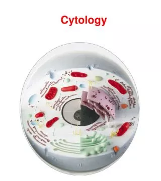
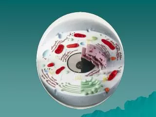
MHD II Laboratory Session Cytology. APRIL 24, 2014. Case 1. Case 1. Q1 Define “cytopathology”. Q2 Define the following terms as they relate to cytopathology and give examples: -“exfoliated cells” -”washings” -”brushings” -”fine needle aspirations”.

E N D
MHD II Laboratory Session Cytology APRIL 24, 2014
Case 1 Q1 Define “cytopathology”
Q2 Define the following terms as they relate to cytopathology and give examples: -“exfoliated cells” -”washings” -”brushings” -”fine needle aspirations”
Q3 What test is currently the most common clinical application of cytopathology?
Case 2 CHIEF COMPLAINT: Annual routine physical examination. HISTORY: The patient is a 31 year-old sexually active female, with a history of several partners. She feels healthy, and has no complaints. PHYSICAL EXAMINATION: Heart, lung, abdominal exams are normal Breast exam is normal and without masses Pelvic exam is unremarkable. A Pap Test is obtained.
Q1For the following patient groups how oftenis a cervical PAP smear indicated? (use the U.S.P.S.T.F. Guidelines – a link is available on LUMEN under “Vertical Curricula”: “Health Maintenance Screening and Prevention” under “Screening Recommendations” • less than 21 years old • 21-29 years old • 30-65 years old • >65 years old • s/p hysterectomy with removal of the cervix and in patients who do not have a history of cervical intraepithelial neoplasia [CIN] grade 2 or 3 or cervical cancer
Q2 Is there a role for testing for Human papillomavirus infection in this patient?
Q3 Describe the steps required to perform a Pap Test (use the images to help your discussion)
Q4 Define “Liquid Based” cytology prep Q5 Define “Conventional” cytology prep
Possible Pap test findings/interpretations in patients are • Normal findings • Evidence of inflammation • Sexually transmitted diseases • Squamous intraepithelial lesion • Carcinoma
Normal cervix cytology/histology Q6 Describe the normal histology Correlate the cytology findings with the histology
Q7 Make a diagnosis based on each of the following PAP smears Your options are: -Evidence of acute inflammation -Herpes simplex virus infection -Trichomonas infection -Human Papillomavirus infection -Squamous cell carcinoma (hints are provided)
C normal
D High Power Correlative tissue section
E hint
Q8 An HPV test is performed in this patient and reveals the presence of a high-risk type of the virus. What is the chance that she will develop a cervical cancer?
Q9 If a woman receives the HPV vaccine does she still need routine Pap Tests?
Case 3 CHIEF COMPLAINT: “I see blood in my urine”. HISTORY: The patient is a 72 year-old-man with a history of low-grade urothelial carcinoma. PHYSICAL EXAMINATION: Physical examination is unremarkable LAB TESTS: Urinalysis – Numerous RBC’s.
Q1 What is the main clinical problem? Q2 Develop a differential diagnosis for this problem.
Diagnostic Work-up Cystoscopy did not reveal any papillary tumors, but did show focal erythematous areas. Bladder washing (barbotage) was performed and the specimen was submitted for cytologic examination.
Q3 Describe the findings in urine specimen (compare to normal, are the cells cytologically benign or malignant?) Normal urothelial cell
The urologist performs a followup bladder biopsy to confirm the diagnosis.
Q4 Describe the histologic findings. Normal bladder
Q6 What is your diagnosis? Q7 What molecular changes can you expect in this patient’s urothelium?
Case 4 CHIEF COMPLAINT: “I found a lump in my neck.” HISTORY: The patient is a 55 year-old-woman with a history of radiation to the neck 20 years ago for hyperthyroidism. PHYSICAL EXAMINATION: A nodule is palpated in the right lobe of the thyroid gland (2 cm) and also an enlarged right cervical lymph node is present. LAB TESTS: Thyroid function tests were normal. Ultrasound confirmed the presence of a 2 cm nodule in the right thyroid and an enlarged lymph node. It was a “cold” nodule on scintiscan.
Q2 A fine needle aspiration (FNA) biopsy is performed. Compare/contrast what is being done in each of the photos below
What are the advantages of FNA? • FNA is SAFE – • Simple, Accurate, Fast and Economic • as well as safe • the best safety record of any method of procuring tissue for a morphologic diagnosis
Describe characteristic cytologic findings. Normal thyroid cytology Q3 What is the cell type present? What structure are the cells forming?
Q4 Patient specimen. What “structure” are the cells forming? Hint – histology correlate
Q6 What is depicted in the image? Hint – histology correlate
Q7 What is your diagnosis? Q8 What molecular changes are associated with this lesion?
Case 5 CHIEF COMPLAINT: “My stomach hurts”. HISTORY: The patient is a 72 year-old woman who presents with vague abdominal pain for the past several months. She denies nausea, vomiting and blood in her stool. PHYSICAL EXAMINATION: The patient is alert and oriented. Vital signs are normal. Her abdominal exam reveals normal bowel sounds with no tenderness to palpation or rebound tenderness. On rectal exam stool is brown and hemoccult negative.
CT scan abdomen CT scan reveals a 4 cm well-demarcated exophytic mass extending into the gastric lumen. Mayo Clinical Proceeding; Volume 83(4), April 2008, p 384
As part of the workup, esophagogastroduodenoscopy was performed, revealing a gastric sub-mucosal mass. This mass was arising from the muscularis propria. Endoscopic ultrasound-guided fine needle aspiration (EUS FNA) was performed. Q2 Briefly describe what an “EUS FNA” is.
Q3. Describe cytologic findings (hint focus on the shape of the cells)
Immunohistochemical stains are performed. C-kit (CD 117)