1 / 13
130 likes | 145 Vues
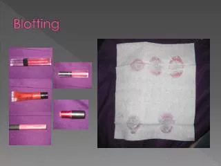
Different blotting techniques are used to identify unique proteins and nucleic acid sequences. Southern, northern, and western blot protocols are similar, and begin with electrophoretic separation of protein and nucleic acid fragments on a gel, which are then transferred to a membrane (nitrocellulose membrane, polyvinylidene difluoride (PVDF) membrane, etc.) where they are immobilized. This enables radiolabeled or enzymatically labeled antibody or DNA probes to bind the immobilized target, and the molecules of interest may then be visualized with various methods. Blotting techniques are select

E N D
Types of blotting Western Blot : The western blot is a widely used analytical technique used to detect specific Proteins in a biological sample Northern Blot : The northern blot is a technique used in molecular biology to study gene expression by detection of RNA (mRNA) in a sample Southern Blot : A Southern blot is a method used in molecular biology for detection of a specific DNA sequence in samples. Southern blotting combines transfer of electrophoresis-separated DNA fragments to a filter membrane and subsequent fragment detection by probe hybridization
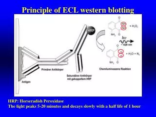
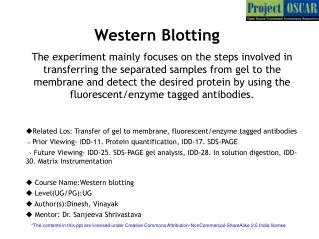
Western blotting • The western blot is a widely used analytical technique used to detect specific proteins in a biological sample. It uses gel electrophoresis to separate native proteins by 3-D structure or denatured proteins by the length of the polypeptide. • The proteins are then transferred to a membrane (typically nitrocellulose), where they are treated with antibodies specific to the target protein. • The gel electrophoresis step is included in western blot analysis to resolve the issue of the cross-reactivity of antibodies. • The proteins will retain or regain part of their structure during blotting that which react with specific antibodies giving rise to the term Immunoblotting.
Electrophoresis Transfer of Proteins to membrane Blocking of proteins Incubation with primary Ab Incubation with secondary Ab Detection & Documentation
Western blotting • The first step is to separate the macromolecules using gel electrophoresis. • Following electrophoresis, the separated molecules are transferred or blotted on a Nitrocellulose or Polyvinylidene Fluoride (PVDF) membrane. • Next, the membrane is blocked to prevent any nonspecific (hydrophobic) binding of antibodies to the surface of the membrane. • The blot is incubated in a dilution of an antiserum i.e, primary antibody directed against the protein of interest. • The transferred protein blot is complexed with an enzyme-labelled secondary antibody as a probe. An appropriate substrate is then added to the enzyme and together they produce a detectable product such as a chromogenic or fluorogenic precipitate on the membrane for colorimetric or fluorometric detection, respectively.
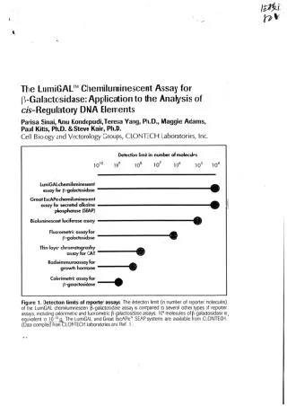
The most sensitive detection methods use a chemiluminescent substrate that, when combined with the enzyme, produces light as a by-product. The light output can be captured using film, a CCD camera or a phosphorimager that is designed for chemiluminescent detection. • Whatever substrate is used, the intensity of the signal correlates with the abundance of the antigen (protein) on the blotting membrane.
Northern blotting • Northern blotting involves the use of electrophoresis to separate RNA samples by size, and detection with a hybridization probe complementary to part of or the entire target sequence. • The northern blot technique was developed in 1977 by James Alwine, David Kemp, and George Stark at Stanford University. Northern blotting takes its name from its similarity to the first blotting technique, the Southern blot, named for biologist Edwin Southern. The major difference is that RNA, rather than DNA, is analyzed in the northern blot. • The term 'northern blot' actually refers specifically to the capillary transfer of RNA from the electrophoresis gel to the blotting membrane. • With northern blotting it is possible to study the structure and function of the particular gene expression rate during differentiation and morphogenesis, in both normal or elevated conditions.
Northern blotting • Advantages of using northern blotting include the detection of RNA size, the observation of alternate splice products, the use of probes with partial homology, the quality and quantity of RNA can be measured on the gel prior to blotting, and the membranes can be stored and reprobe for years after blotting. • Blot Base is an online database publishing northern blots. Blot Base has over 700 published northern blots of human and mouse samples, in over 650 genes across more than 25 different tissue types. Northern blots can be searched by a blot ID, paper reference, gene identifier, or by tissue. The results of a search provide the blot ID, species, tissue, gene, expression level, blot image (if available), and links to the publication that the work originated from. • Northern blotting allows to observe gene expression patterns between tissues, organs, developmental stages, environmental stress levels, pathogen infection, and over the course of treatment. • The technique has been used to show over expression of oncogenes and down regulation of tumor-suppressor genes in cancerous cells when compared to 'normal' tissue. • A problem in northern blotting is often sample degradation by RNases which can be avoided by proper sterilization of glassware and the use of RNase inhibitors such as DEPC (diethylpyrocarbonate). • The chemicals used in most northern blots can be a risk-since formaldehyde, radioactive material, ethidiumbromide, DEPC, and UV light are all harmful under certain exposure
If you would like to donate us? Scan below and donate us 0.013$ (US dollar) (5Rs Indian rupee) Contact: If you want PPT/PDF files, please contact below. Email: gnccmysore@gmail.com Telegram:+919738137533(only for Chat)