Download
1 / 40
440 likes | 797 Vues
Southern, Northern and Western blotting. 生理所 黃阿敏. Comparison of Southern, Northern, and Western analyses of Gene X. Southern hybridization. First described by E. M. Southern in 1975. Applications of Southern hybridization RFLP ’ s, VNTR ’ s and DNA fingerprinting

E N D
Comparison of Southern, Northern, and Western analyses of Gene X
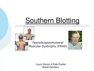
Southern hybridization • First described by E. M. Southern in 1975. • Applications of Southern hybridization • RFLP’s, VNTR’s and DNA fingerprinting • Checking of the gene knockout mice • The flow chart of Southern hybridization
Southern hybridization Transfer buffer
Detection of the sickle-cell globin gene by Southern blotting
Flow chart of Southern hybridization Preparing the samples and running the gel Southern transfer Probe preparation Prehybridization Hybridization Post-hybridization washing Signal detection Isotope Non-isotope
Preparing the samples and running the gel • Digest 10 pg to 10 g of desired DNA samples to completion. • Prepare an agarose gel, load samples (remember marker), and electrophorese. • Stain gel ethidium bromide solution (0.5 g/ml). • Photograph gel (with ruler).
Critical parameters (I) • Note the complexity of DNA • Genomic DNA • A single-copy of mammalian gene, 3 Kb average in length 10 mg x 3 Kb/3 x 106 Kb = 10 mg x 1/106 = 10 pg • Plasmid DNA or PCR products 0.1 mg of a 3 Kb plasmid DNA 100 ng
Gel treatment • Acid treatment • 0.2 N HCl solution • Denaturation • NaOH solution • Neutralization • Tris-Cl buffer (pH8.0)
Southern transfer • Measure gel and set up transfer assembly: • Wick in tray with 20x SSC • Gel • Nitrocellulose or Nylon filters (soaked in H2O and 20x SSC) • 3MM Whatman filter paper • Paper towels • Weight
After Southern transfer • Dissemble transfer pyramid and rinse nitrocellulose in 2x SSC • Bake nitrocellulose at 80C for 2 hr or UV-crosslink Nylon membrane for seconds
Preparation of probes • Synthesis of uniformly labeled double-stranded DNA probes • Preparation of single-stranded probes • Labeling the 5 and 3 termini of DNA
Synthesis of double-stranded DNA probes • Nick translation of DNA • Labeled DNA probes using random oligonucleotide primers
Preparation of single-stranded probes • Synthesis of single-stranded DNA probes using bacteriophage M13 vectors. • Synthesis of RNA probes by in vitro transcription by bacteriophage DNA-dependent RNA polymerase.
Labeling the 3 termini of double-stranded DNA using the Klenow fragment of E. coli DNA polymerase I. (lack of 5’ 3’ exonuclease activity) Labeling the 3 termini of double-stranded DNA using bacteriophage T4 DNA polymerase. Labeling the 5 termini of DNA with bacteriophage T4 polynucleotide kinase. Labeling the 5 and 3 termini of DNA
Non-isotope labeling • Digoxigenin-11-dUTP (DIG-dUTP) labeling • DNA labeling • Oligonucleotide labeling • RNA labeling
Prehybridization • Add prehybridization solution and prehybridize at hybridization temperature for 2-4 hr
Hybridization • Remove prehybridization solution and add hybridization solution • Add 500,000 cpm of the probe/ml hybridization solution. • Hybridize overnight at appropriate temperature.
Post-hybridization washing • Wash twice, 15 min each, in 1x SSC, 0.1% SDS at room temperature. • Wash twice, 15 min each, in 0.25x SSC, 0.1%SDS at hybridization temp
Critical parameters (II) • Homology between the probe and the sequences being detected • Tm = 81 +16.6 (log Ci) + 0.4 [% (G+C)] - 0.6 (% formamide)- 600/n - 1.5 (% mismatch) • Factors can be changed: • Hybridization temp. • Washing temp. • Salt concentration during washing High temp., low salt: high stringency Low temp., high salt: low stringency • If 50 % formamide is used • 42 oC for 95 ~ 100 % homology • 37 oC for 90 ~ 95 % homology • 32 oC for 85 ~ 90 % homology
Signals detection • Autoradioragraphy • Non-isotope detection system • Chemiluminescent detection • Colorimetric detection • Multicolor detection
Autoradiography • Exposure to x-ray film
Northern blotting or Northern hybridization • Technique for detecting specific RNAs separated by electrophoresis by hybridization to a labeled DNA probe.
The flow chart of Northern hybridization Prepare RNA samples and run RNA gel Northern transfer Probe preparation Prehybridization Hybridization Post-hybridization washing Signal detection Isotope Non-isotope
Preparation of agarose/formaldehyde gel • E.g. Prepare a 350 ml 1.2% agarose/formaldehyde gel • 4.2 g agarose in 304.5 g water. Microwave, then cool to 60C. Add 35 ml 10x MOPS running buffer and 10.5 ml 37% formaldehyde
Preparation of RNA samples • Prepare a premix: • 5 l of 10x MOPS running buffer • 8.75 l of 37% formaldehyde • 25 l of formamide. • Prepare RNA samples: • 38.75 l of premix • RNA (0.5 to 10 g)* • water to 50 l • *If the mRNA species of interest makes up a relatively high percentage of the mRNA in the cell (>0.05% of the message), total cellular RNA can be used. If the mRNA species of interest is relatively rare, however, it is advisable to use poly(A)+ RNA. • Incubate 15 min at 55C
Running the RNA gel • Add 10 l formaldehyde loading buffer to each sample and load gel. Run gel at 100 to 120 V for ~3hr. • Remove gel from the running tank and rinse several times in water. Place gel in 10x SSC for 45 min. • Do not need post-transferring gel treatment
An example of Northern blotting Northern blot 28 S RNA gel 18 S
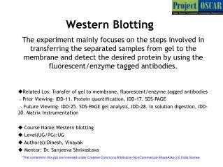
Western blotting, or immunoblotting Technique for detecting specific proteins separated by electrophoresis by use of labeled antibodies.
Flow chart of Western blotting Electrophoresing the protein sample Assembling the Western blot sandwich Transferring proteins from gel to nitrocellulose paper Staining of transferred proteins Blocking nonspecific antibody sites on the nitrocellulose paper Probing electroblotted proteins with primary antibody Washing away nonspecifically bound primary antibody Detecting bound antibody by horseradish peroxidase-anti-Ig conjugate and formation of a diaminobenzidine (DAB) precipitate Photographing the immunoblot
Analysis of protein samples by SDS polyacrylamide-gel electrophoresis and Western blotting Protein bands detected by specific antibody SDS-PAGE Western blot
Comparison of Southern, Northern, and Western blotting techniques