Download
1 / 114
1.15k likes | 1.58k Vues
Mechanisms of protection against infection and disease are diverse. Primarily they can be divided into two major categories: non-specific (innate) and specific (adaptive), which differ as follows : .

E N D
Mechanisms of protection against infection and disease are diverse. Primarily they can be divided into two major categories: non-specific (innate) and specific (adaptive), which differ as follows:
The elements of the innate immune system: anatomical barriers, secretory molecules, cellular components. Anatomical barriers:Skin, intestinal movement, oscillation of broncho-pulmonary cilia, etc. Prevent pathogens from entering and/or getting a foothold in the body.
Secretory molecules: organic acids in skin secretions, lysozyme in oro-naso-pharyngeallacrimal secretions (Of or relating to tears), thiocyanate in saliva,low molecular weight fatty acids in the lower bowel; bile acids and low molecular weight fatty acids in lower GI tract; transferrin,lactoferrin, lysozyme, interferons, fibronectin, complement, acute phase proteins, etc. in serum; Interferons and tumor necrosis factor (TNF) at the site of inflammation.
Transferrin and lactoferrin deprive organisms of iron. Interferons inhibit viral replication and activate other cells which kill pathogens. lysozyme, in serum and tears, breaks down the bacterial cell wall (peptidoglycan); fibronectin coats (opsonizes) bacteria and promotes their rapid hagocytosis.
Complement components and their products cause destruction of microorganism directly or with the help of phagocytic cells. Acute phase proteins (such as CRP) interact with the complement system proteins to combat infections.TNF-a up presses viral replication and activates phagocytes (nitric oxide pathway).
Cellular Components: Phagocytic cells: Neutrophils (PMN) ,macrophages and monocytes are the most important cellular components of the non-specific immune system. Neutrophils : are most important cellular components in bacterial destruction.
They are relatively large and most abundant white blood cells with lobed nucleus and cytoplasmic granules (lysosomes). They are identified by their characteristic morphology. In addition, monoclonal antibodies against characteristic cell surface protein, cluster differentiation marker, CD66 can be used to identify these cells.
PMN granules are of two kinds: primary (azurophilic) and secondary (specific). Primary azurophilic granules are characteristic of immature and very young neutrophils. They contain NADPH oxidase co-factors, cationic proteins, defensins (small molecular weight proteins),proteases (elastase, cathepsin G, etc.), lysozyme and myeloperoxidase (characteristic of primary granules).
Secondary granules are more characteristic of (specific for) mature neutrophils. They contain lysozyme and NADPH oxidase cofactors, lactoferrin and B-12-binding protein, the last two are characteristic for these granules.
Mononuclear phagocytes are the other population of phagocytic cells and include monocytes in circulation, histiocytes in tissues, microglilal cells in the brain, Kupffer cells in the liver and MQs in serous cavities and lymphoid organs. They also have granules similar to those in neutrophils, although not as abundant. They are recognized by their morphology, ability to adhere on glass/plastic surface, phagocytic property and CD14 marker.
All phagocytic cells have receptors for a variety of molecules.Most pertinent to non-specific immunity are scavenger and TOLL-like receptors and receptors for the Fc part of IgG, complement, interferons, TNF and certain bacterial components.Interaction of organisms with these receptors promotes phagocytosis and phagocyte activation for a more efficient killing of pathogens.
Phagocyte response to infection: Chemotaxis: Bacteria produce N-formyl-methionine-containing peptides which are powerful attractants (chemotactic) for phagocytic cells. Many bacteria also act on proteins of the complement and clotting systems to produce peptides that cause vasodialation vascular permeability and expression of adherence molecules on vascular endothelial cells.
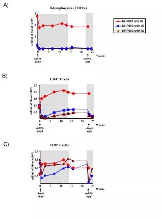
Classes of Iymphocytes B lymphocytes recognize soluble antigens and develop into antibody-secreting cells Helper T lymphocyte recognize antigens on the surfaces of APCs and secrete cytokines, which stimulate different mechanisms of immunity and inflammation CTLs recognize antigens on infected cells and kill these cells Regulatory T cells suppress and prevent immune response, e.g. to self antigens NK cells use receptors with more limited diversity than T or B cell antigen receptors to recognize and kill their targets, such as infected cells
The Adaptive Immune Respons The adaptive immune system uses three main strategies to combat most microbes. . Secreted antibodies bind to extracellularmicrobes, block their ability to infect host cells, and promote their ingestion and subsequent destruction by phagocytes. . Phagocytes ingest microbes and kill them, and helper T cells enhance the microbicidal abilities of the phagocytes. . CTLs destroy cells infected by microbes that are inaccessible to antibodies.
Phases of adaptive immune responses Adaptive immune responses consist of distinct phases, the first three being the recognition of antigen. the activation of lymphocytes. and the elimination of antigen (the effector phase). The response contracts (declines) as antigen-stimulated lymphocytes die by apoptosis. restoring homeostasis, and the antigen-specific cells that survive are responsible for memory. The duration of each phase may vary in different immune responses. The y-axis represents an arbitrary measure of the magnitudeof the response. These principles apply to humoral immunity (mediated by B lymphocytes) and cell-mediated immunity (mediated by Tlymphocytes).
They also induce, on phagocytic cells, expression of proteins (e.g., integrins) that promote binding to endothelial cells. Phagocytic cells respond to chemotactic peptides of bacterial and host origin and migrate across the capillary wall (diapedesis) to the site of infection/inflammation.
Attachment: Once at the site of infection, phagocytes can attach to bacteria via a number of receptors for bacterial components (scavenger receptor, LPS receptor, mannose receptor, TOLL-like receptors, etc.) or host proteins (opsonins) that coat (opsonize) bacteria (e.g., fibronectin, complement, IgG antibody, etc.).
This attachment triggers the activation of respiratory burst (hexose monophosphate shunt), internalization of the organisms (phagosome formation), phagosome lysosome fusion (phagolysosome) etc.
Respiratory burst and phagocytosis: The process of attachment and phagosome formation is accompanied by the activation of the respiratory burst (hexose monophosphate shunt) which results in the production of superoxide anion, singlet oxygen, hydroxyl ion and hydrogen peroxide . These molecules are microbicidal and cause killing of organisms in the phagosome.
Phagosome-lysosome fusion: Phagosome, soon after its formation, fuses with granules (lysosomes) to form a phago-lysosome. As mentioned earlier, lysosomes contain a variety of anti-microbial substances (e.g., lysozyme, defensins, proteases, lactoferrin, transferrin,etc.) and phago-lysosome fusion results in the exposure of microorganisms to these substances and their destruction.
Also, fusion of phagosome with primary granules (in newly recruited phagocytes) that contain myeloperoxidase results in the production of OCl , (or other halides: I and Br) and halogenation of bacterial proteins. This ultimately leads to bacterial killing.
Three modes of intracellular killing by phagocytosis:(1) by lysosomal antibacterial substances (lactoferrin, cationic proteins, lysozyme, defensins, proteases, etc.) without the requirement of respiratory burst (oxygen-independent killing.
(2) by products of respiratory burst (super-oxide, singlet oxygen, hydroxyl radical, hydrogen peroxide, etc.) without the need for myeloperoxidase (oxygen-dependent, myeloperoxidase-independent killing. (3) by halogenation of bacterial proteins catalyzed by meyloperoxidase (oxygen-dependent, myeloperoxidase-dependent killing:
These processes occur simultaneously and act synergistically. A defect in any of thesepathways, for example, a deficiency of NADPH oxidase (cytochrome b558) components (p91, p22,47 p61-phox), myeloperoxidase, etc. would impair the killing activity of phagocytes and render the host more susceptible to pyogenic infections. NADPH oxidase is the most serious of all these defects.
Neutrophils also contain catalase and glutathion (GS) which detoxify excess H2O2 . GS, in its reduced form (GSH), also recycles NADP to NADPH. Interaction of phagocytic cells with certain humoral factors (e.g. interferons, TNF, , etc.) can increase their phagocytic function,respiratory burst and intra-cellular killing.Certain proteins secreted by various cells (cytokines) can also induce phagocytic cells, particularly macrophages, to produce nitric oxide (NO) that is toxic to microorganism and malignant cells.
آنتی ژن Agهر ماده ای که بتواند با پاسخ ایجاد شده توسط سیستم ایمنی واکنش اختصاصي نشان دهد آنتی ژن نام دارد یا اینکه بتواند خود موجب ایجاد پاسخ ایمنی گردد . آنتي ژن ريشه يوناني دارد : Gen= producing وAnti=against بطور کلی آنتی ژن ها دو دسته هستند :1-توانایی تحریک سیستم ایمنی را دارند و پاسخ سیستم را به راه می اندازند به عبارتی موجب تحریک و پاسخ می شوند که به این دسته ایمونوژن Immunogene گفته می شود.- دسته ای که توانایی تحریک و به راه افتادن پاسخ سیستم را ندارند به آن ها هپتن Haptenمی گویند.یکی از دلایل عدم تاثیر هاپتن ها بر روی سیستم ایمنی مربوط به کوچکی اندازه مولوکول است لذا برای رفع این نقیصه Hapten ها را به موادی به نام حامل یا Carrir اتصال می دهد که در نتیجه تحریک و پاسخ را سبب می شود .تذکر هر آنتی ژنی ایمنوژن نیست ولی هر ایمنوژنی آنتی ژن می باشد.
دسته اي از آنتي ژنهاسوپر آنتي ژن نام دارند كه بر عكس آنتي ژنهاي معمولي بدون نياز به عرضه آنتي ژن توسط ماكروفاژها، مستقيما از طريق مجموعه آنتي ژنهاي سطح MHC II در اين سلولها، لنفوسيتهاي T را با اتصال به ناحيه متغير زنجيره بتاي واريابل گيرنده (vβ) آنتي ژني تحريك مي كنند.آنتي ژنهاي معمولي از هر 10000 تا 100000 T Cell يكي را تحريك مي كنند در حاليكه سوپر انتي ژنها از هر 5 تا 50 يكي را تحريك مي كنند، لذا سوپر آنتي آنتي ژنها به سرعت و در مدت 3 ساعت تعداد زيادي از T Cell ها را تحريك كرده و منجر به ترشح مقدار زيادي اينترلوكين- 2((IL-2 ، اينترفرون گاما ((INF-γ و فاكتور نكروز دهنده تومور (TNF) مي گردد.ترشح زياد اين مواد در بدن مي تواند تظاهرات مسموميت غذايي، تب شديد و در مواردي سبب مرگ شود. تعدادي از سوپر آنتي ژنها به شرح زير است:
Staphylococcus centrotoxinsToxic Shock Syndrome Toxin-1(TSST-1),(staphylococcal exotoxin)Staphylococcal exfolitative toxin or epidermolytic toxinStroptococcal exotoxin-AStroptococcal M proteinsRabies nucleucapsid proteinMycoplasma arthritidisMurine leukemia virus(MuL-v)HIV virus (gp 120?)ایمنوژنیسیته : به معني قدرت ايمني زايي يك ماده ، خاصیتی که ایمونوژن برای تحریک سیستم ایمنی از خود نشان می دهد است.
ویژگی های یک مولکول برای ایمنوژنیسیته بودن :ویژگی های آنتی ژن و 2- ویژگی های میزبانویژگی های آنتی ژن1- اندازه مولکولی Molecular sizeیک آنتی ژن اندازه های مختلفی می تواند داشته باشد و هر چه این اندازه بزرگتر باشد آنتی ژنیسیتی آن بیشتر است. مولکول های دارای اندازه ی مولکولی -100000-10000 دالتون باشد قدرت آنتی ژنیسیتی متوسط دارد.ميكروبها، ويروسها و گلبولهاي قرمز با وزن مولكولي بالاتر از 100000 دالتون قدرت آنتي ژني قوي دارند ولي از 10000 به پایین قدرت آنتي ژني کمتر بوده واصلا آنتی ژن محسوب نمی شود.
2- پیچیدگی Complexity معمولاَ آنتی ژن هر چه از لحاظ ساختمانی پیچیده تر باشد از نظر آنتی ژنیسیتی قویتر است و این مسئله تا حدودی به ماهیت شیمیائی آنتی ژن ها بستگی دارد مثلا" پروتئین ها چون از اسید آمینه هایی با تنوع زیاد تشکیل شده اند لذا می توان مولکول های پیچیده ای ایجاد کرده و قدرت آنتيژني بالاتری داشته باشند.آندسته از مولکول هایی که دارای اسید آمینه آروماتیک باشند از لحاظ قدرت آنتی ژن قویترند مانند تیروزین یا فنیل آلانین.پلی ساکارید ها به دلیل ساختمان یکسانشان، هموپليمر،(و لیپید ها و اسید های نوکلئیک ) از لحاظ آنتی ژنبک ضعیف هستند و در صورتی که با پروتئین ها ترکیب شوند قدرت آن ها بالا می رود
3-ثبات ساختمانی Stability یک آنتی ژن وقتی قوی محسوب می شود که ثبات ساختمانی خوبی داشته باشد ،این ثبات با قدرت تحریک یا Flexibility متفاوت است مثلاً فلاژلین که در فلاژل باکتری ها بوده و دارای قدرت تحریک زیادی است از نظر ثبات ساختمانی ضعیف بوده و لذا از لحاظ آنتی ژنیک هم ضعیف است . 4- قدرت تجزیه شدن Degradebility بسته به نوع پاسخ ایمنی وضعیت آنتی ژن متفاوت است بطوریکه در پاسخ ایمنی همورال لنفوسیت های B آنتی ژن را در حال سه بعدی شناسائی می کنند ولی لنفوسیت های T هنگامی آنتی ژن را شناسائی می کنند که قبلاً توسط سلول های عرضه کننده آنتی ژن پروسس شده باشند.
5- بیگانگی آنتی ژن Foriegnness هر چقدر آنتی ژن نسبت به بدن غیر خودی باشد non self پاسخ به آن نیز سریعتر می باشد . تجربیات نشان میدهد در پیوند عضو بین دوقلوهای متشابه مونوزیگوت پیوند رفع نمی شود اما در دوقلوهای هتروزیگوت احتمال دفع زیاد تر است و هرچقدر فاصله خویشاوندی بیشتر باشد ،شدت پاسخ برای دفع بیشتر است مثلاً پیوند کلیۀ شمپانزه به انسان بیشترین پاسخ را ایجاد می کند. 6- مسیرهای تجویز آنتی ژن در حالیکه یک آنتی ژن ممکن است وقتی داخل رگی تزریق شود تولید پاسخ نکند اما تزريق زير پوستي آن ايجاد پاسخ مناسبي بكند. 7- شکل فضایی آنتی ژن Conformation هر چه شکل فضایی مولکول بیشتر پیچیده باشد و دارای زنجیره های جانبی فراوان تری باشد قدرت آنتی ژنیسیتی آن هم بیشتر است .شاخصهاي شكلي قدرت ايمني زايي بيشتري دارند نسبت به ساير شاخصهاي يك آنتي ژن.اين شاخصها بيشتر در زواياي مولكول قرار دارند و در نتيجه قدرت چرخش ((Flexibilityزيادي دارند و بهتر مي توانند سيستم ايمني را تحريك نمايند.
8-بار الكتريكي آنتي ژن (Charges) بار الكتريكي هر آنتي ژن در قدرت ايمني زايي و خصوصيات آن آنتي ژن نقش دارد.از آنجايي كه بارهاي الكتريكي هيدروفيلي آنتي ژن باعث كشش و تماس بيشتري بين مولكولها مي شوند، بنا بر اين بار الكتريكي و در دسترس بودن اپي توپ هاي آنتي ژن در تحريك سيستم ايمني بدن لازم و ملزوم يكديگرند. بطور استثناء مولكولهايي كه كاملا فاقد بار الكتريكي هستند ممكن است در مواردي ايمونوژنيك شوند مانند دكستران (.(Dextran
بار الكتريكي مطلق ((Net charge يك آنتي ژن با آنتي بادي ضد آن نسبت عكس دارد مثلا پروتئين با بار مثبت در بدن حيوان آزمايشگاهي آنتي بادي با بار الكتريكي مطلق منفي توليد مي كند. اتصال هاپتن به اين پروتئين تغييري در بار الكتريكي مطلق آنتي بادي عليه آن ايجاد نمي كند. 9- Optical configuration چرخش نوري اسيدهاي آمينه موجود در طبيعت اكثرا از نوعَ L-Amino Acid هستند و آنزيمهايي كه در بدن هستند اين دسته پروتئينها را متابوليزه كرده و عليه آنها عكس العمل نشان مي دهند. پلي پپتيدهاي مصنوعي با اسيد آمينه هاي ِD اگر به حيوان تزريق شود ايجاد پاسخ نخواهد كرد. اگر مولكولهاي يك هاپتن مثل پني سيلين را به پليمر اسيد آمينه هاي نوع D اسيد گلوتاميك و -D ليزين پيوند دهيم و به موش تزريق شود ايمني موش عليه پني سيلين مهار مي شود كه (Immunological tolerance) نسبت به پني سيلين كه به يك حامل ايمونوژن متصل شده است ايجاد شده است.
10-مقدار آنتي ژن مقدار آنتي ژن تزريقي در نوع پاسخ ايمني عليه آن انتي ژنها نقش مهمي دارد.مقدار كم آنتي ژن بتدريج يا مقادير زياد يكباره اگر تزريق شود ممكن است تحمل ايجاد شود. روش قديمي بسردكا بر همين مبنا بوده و سرم همولوگ حيواني را براي درمان يك بيماري كه به آن سرم حساس است و نجات وي با آن سرم ممكن مي شود را تزريق مي كنند.امروزه به آن حساسيت زدايي يا Desensitization گويند. براي درمان ايمونولوژيكي بيماريهاي آلرژي آتوپي ((Atopyَ هم از اين روش استفاده مي شود. Optimal dose يك آنتي ژنبهترين پاسخ را ايجاد مي كند.