Download

1 / 20
220 likes | 504 Vues
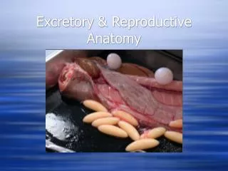
By C. Kohn, Waterford, WI. Bovine Reproductive Anatomy. BASIC ANATOMY OF THE COWS REPRODUCTIVE SYSTEM. The cow's reproductive system has four basic functions. To produce ova (eggs) which provides half of the eventual offspring's genetic makeup.

E N D
By C. Kohn, Waterford, WI Bovine Reproductive Anatomy
BASIC ANATOMY OF THE COWS REPRODUCTIVE SYSTEM • The cow's reproductive system has four basic functions. • To produce ova (eggs) which provides half of the eventual offspring's genetic makeup. • To provide an environment and conditions for the fertilization of those ova. • To provide a place following fertilization for the nourishment and fetal development of the calf. • To provide a mechanism for the birth of the calf. ~
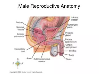
Female Repro Structures • Vulva • Vagina • Cervix • Uterus • Oviducts & Infundibulum • Ovaries • Corpus Luteum • Follicles & Eggs ~ Look at this picture & predict structure function
Vulva • “Entranceway” of the female reproductive tract • Only part visible from the outside • Swells and becomes reddish-pink during estrus • Response due to estrogen ~
Vagina • Vagina – flattened tube; passage between the cervix and the vulva • Site of semen deposition during natural insemination • Used as passageway for instruments during AI • Produces mucus (lubricant)- flushes out irritants and infectious agents • Common site of infection ~
Cervix • Cervix – the muscular “valve” or “control gate” between the uterus and the vagina • Made of muscular folds that slow down invading materials • These folds have ‘dead ends’ that trap foreign substances • Completely closed except during estrus and parturition (calving) • During pregnancy, a hard mucus plug “glues” it shut ~
Uterus & Uterine Horns • Uterus – where the fetus grows, a.k.a. womb • Muscular, capable of “enormous expansion” • Has to support up to 80 kg / 177 lbs of weight • Uterine Horns • The extensions on either side of the uterus that lead to the oviducts • Curl like ram horns ~
Oviducts (Fallopian Tubes) • Oviducts– tubes that carry eggs from ovaries to uterus • Kept shut tight except during ovulation and insemination • Where fertilization occurs • Egg moved from the ovaries down the oviduct by cilia (microscopic hairs) • Motile sperm meet the egg in the upper part of the oviduct • Newly formed zygote stays in the oviduct 3-4 days • This time is needed for the uterus to prepare itself ~
Infundibulum • Infundibulum – Latin for “funnel” • The end projection of the oviducts that surrounds, but does not connect to, the ovaries • “Funnels” eggs from ovaries into oviduct. ~
Ovaries • Small walnut-shaped ovals 4-6 cm / 2-3 inches in length • Contain thousands of ova (plural of ovum, or egg cell) • These were created before the birth of the cow • Has a finite supply, as do human females ~
Ovaries (cont.) • Functions: • Produce a mature ovum (egg) every 21 days • Produce/secrete hormones that: • Control growth of egg • Change cow’s behavior (gets her “in the mood”) • Prepare reproductive tract for pregnancy • Start parturition process (birthing) • Prepare mammary glands for lactation ~
Follicles • Follicles – start as cavities (holes) on the ovary • An egg moves to this cavity. • It is surrounded by support cells and nutritive substances • All these things together are the follicle ~
Corpus luteum • The cells that remain in the follicle after the egg is ovulated (expelled into the oviduct) become the corpus luteum (CL) • Corpus luteum translated = yellow body • Produces progesterone, a hormone which sustains the pregnancy (allows pregnancy to “progress”) • Occurs regardless of fertilization ~
Egg (ovum) • Female gamete (reproductive cell) • Haploid - half the number of normal chromosomes • Present prior to birth, but maturation occurs at puberty • Multiple eggs develop during a cycle, but only one matures ~
This is an animation – to watch the animation, use a computer or laptop.
How they change during estrus • Vulva: swollen due to estrogen, covered in mucus • Vagina: excess mucus production • Cervix: dilates to allow acceptance of semen (otherwise locked shut with hardened mucus to prevent infection) • Oviducts: open to allow ovulation, fertilization • Ovaries: ovulation – release of the follicle (egg and some supporting cells) from the ovary • number of young that a female can produce at one time is determined by how many eggs are released during ovulation • ovulation usually occurs at the end of a heat/estrus ~ MAKE SURE YOU KNOW THIS!
Anatomical Disorders • Closed Cervix – cervix does not open to allow fert. • Retained Placenta – afterbirth stays in cow • Damaged Oviduct (due to excess palpation) • Freemartins – heifer exposed to male hormones • Cystic ovaries – growth/swelling of ovaries • Infection – varies • Anovulation – lack of ovulation • Metritis – inflammation of lining of the uterus ~