REPRODUCTIVE ANATOMY
REPRODUCTIVE ANATOMY. FEMALE REPRODUCTIVE SYSTEM. The female reproductive system is designed to carry out several functions: produces the female egg cells necessary for reproduction called the Ova designed to transport the ova to the site of fertilization

REPRODUCTIVE ANATOMY
E N D
Presentation Transcript
FEMALE REPRODUCTIVE SYSTEM • The female reproductive system is designed to carry out several functions: • produces the female egg cells necessary for reproduction called the Ova • designed to transport the ova to the site of fertilization • Conception, the fertilization of an egg by a sperm, normally occurs in the fallopian tubes. • The next step for the fertilized egg is to implant into the walls of the uterus, beginning the initial stages of pregnancy. • If fertilization and/or implantation does not take place, the system is designed to menstruate (the monthly shedding of the uterine lining). • The female reproductive system produces female sex hormones that maintain the reproductive cycle.
FEMALE REPRODUCTIVE SYSTEM • The PRIMARY function of the external female reproductive structures (the genitals) is twofold: • (1)To enable sperm to enter the body • (2) to protect the internal genital organs from infectious organisms.
FEMALE REPRODUCTIVE ANATOMYEXTERNAL STRUCTURES • Labia majora:enclose and protect the other external reproductive organs. Comparable to the scrotum in males. Contain sweat and oil-secreting glands. After puberty, are covered with hair. Translated as “large lips.” • Labia minora: can be very small or up to 2 inches wide. They lie just inside the labia majora, and surround the openings to the vagina (the canal that joins the lower part of the uterus to the outside of the body) and urethra (the tube that carries urine from the bladder to the outside of the body). translated as "small lips • Bartholin's glands: These glands are located beside the vaginal opening and produce a fluid (mucus) secretion. • Clitoris: The two labia minora meet at the clitoris, a small, sensitive protrusion that is comparable to the penis in males. The clitoris is covered by a fold of skin, called the prepuce, which is similar to the foreskin at the end of the penis. Like the penis, the clitoris is very sensitive to stimulation and can become erect.
FEMALE REPRODUCTIVE ANATOMY INTERNAL STRUCTURES • Vagina:a canal that joins the cervix (the lower part of uterus) to the outside of the body. It also is known as the birth canal. • Uterus (womb):a hollow, pear-shaped organ that is the home to a developing fetus. Divided into two parts: the cervix, which is the lower part that opens into the vagina, and the main body of the uterus, called the corpus. The corpus can easily expand to hold a developing baby. A channel through the cervix allows sperm to enter and menstrual blood to exit. • Ovaries:small, oval-shaped glands that are located on either side of the uterus. The ovaries produce eggs and hormones. • Fallopian tubes: These are narrow tubes that are attached to the upper part of the uterus and serve as tunnels for the ova (egg cells) to travel from the ovaries to the uterus. Conception normally occurs in the fallopian tubes. The fertilized egg then moves to the uterus, where it implants into the lining of the uterine wall.
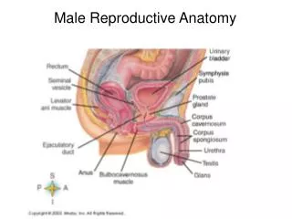
MALE REPRODUCTIVE SYSTEM • The purpose of the organs of the male reproductive system is to perform the following functions: • To produce, maintain, and transport sperm (the male reproductive cells) and protective fluid (semen) • To discharge sperm within the female reproductive tract during sex • To produce and secrete male sex hormones responsible for maintaining the male reproductive system
MALE REPRODUCTIVE SYSTEM • Most of the male reproductive system is located outside of the body. These external structures include: • the penis • Scrotum • testicles.
MALE REPRODUCTIVE ANATOMYEXTERNAL STRUCTURES • Penis: Organ used in sexual intercourse. It has three parts: • the root, which attaches to the wall of the abdomen; • the body, or shaft; • the glans, which is the cone-shaped part at the end of the penis. The glans, also called the head of the penis, is covered with a loose layer of skin called foreskin. • The opening of the urethra, the tube that transports semen and urine, is at the tip of the penis. • The body consists of three circular shaped chambers. These chambers are made up of special, sponge-like tissue. This tissue contains thousands of large spaces that fill with blood when the man is sexually aroused. As the penis fills with blood, it becomes rigid and erect, which allows for penetration during sexual intercourse. The skin of the penis is loose and elastic to accommodate changes in penis size during an erection. • Semen, which contains sperm (reproductive cells), is expelled (ejaculated) through the end of the penis when the man reaches sexual climax (orgasm). When the penis is erect, the flow of urine is blocked from the urethra, allowing only semen to be ejaculated at orgasm.
MALE REPRODUCTIVE ANATOMY EXTERNAL STRUCTURES • Scrotum: This is the loose pouch-like sac of skin that hangs behind and below the penis. It contains the testicles. • The scrotum acts as a "climate control system" for the testes. For normal sperm development, the testes must be at a temperature slightly cooler than body temperature. Special muscles in the wall of the scrotum allow it to contract and relax, moving the testicles closer to the body for warmth or farther away from the body to cool the temperature.
MALE REPRODUCTIVE ANATOMY EXTERNAL STRUCTURES • Testicles (testes): These are oval organs about the size of large olives that lie in the scrotum, secured at either end by a structure called the spermatic cord. • Most men have two testes. • The testes are responsible for making testosterone, the primary male sex hormone, and for generating sperm. • Within the testes are coiled masses of tubes called seminiferous tubules. These tubes are responsible for producing sperm cells.
MALE REPRODUCTIVE ANATOMY INTERNAL STRUCTURES • Epididymis:a long, coiled tube that rests on the backside of each testicle. It transports and stores sperm cells that are produced in the testes. It also is the job of the epididymis to bring the sperm to maturity. During sexual arousal, contractions force the sperm into the vas deferens. • Vas deferens:a long, muscular tube that travels from the epididymis into the pelvic cavity, to just behind the bladder. Transports mature sperm to the urethra in preparation for ejaculation. • Ejaculatory ducts: These are formed by the fusion of the vas deferens and the seminal vesicles. The ejaculatory ducts empty into the urethra
MALE REPRODUCTIVE ANATOMY INTERNAL STRUCTURES • Urethra:the tube that carries urine from the bladder to outside of the body. It has the additional function of ejaculating semen when the man reaches orgasm • Seminal vesicles:sac-like pouches that attach to the vas deferens near the base of the bladder. Produce a sugar-rich fluid (fructose) that provides sperm with a source of energy to help them move. The fluid of the seminal vesicles makes up most of the volume of a man's ejaculatory fluid, or ejaculate. • Prostate gland:a walnut-sized structure that is located below the urinary bladder in front of the rectum. Contributes additional fluid to the ejaculate. Prostate fluids also help to nourish the sperm. The urethra, runs through the center of the prostate gland. • Bulbourethral glands: Also called Cowper's glands, these are pea-sized structures located on the sides of the urethra just below the prostate gland. Produce a clear, slippery fluid that empties directly into the urethra. This fluid serves to lubricate the urethra and to neutralize any acidity that may be present due to residual drops of urine in the urethra.