Download
1 / 38
390 likes | 676 Vues
Development of Reproductive Anatomy. Development of sex organs: overview All the same, until week 7 – then differentiation The internal genitalia The external genitalia Problems Oogenesis. Development of sex organs: overview.

E N D
Development of Reproductive Anatomy • Development of sex organs: overview • All the same, until week 7 – then differentiation • Theinternal genitalia • The external genitalia • Problems • Oogenesis
Development of sex organs: overview • Sex determination occurs first in the gonads and is followed by appropriate development of the ducts, glands and associated genitalia. • In the absence of testis-determining genes, the gonadal primordium develops as an ovary
I. All the same, until week 7 Sexually indifferent stage: By 6-7 weeks of fetal life, fetuses of both sexes have two sets of internal ducts, the Mullerian (female) ducts and the Wolffian (male) ducts. • Gonad differentiation: female – ovaries
Mammalian gonad forms within the developing urogenital system, which itself derives from the intermediate mesoderm. Epithelial structures are shown in red, mesenchymal structures are shown in blue, and the striped region denotes the genital ridge. (WD) Wolffian duct; (MT) mesonephric tubules; (MD) Mullerian duct; (UB) ureteric bud; (CE) coelomic epithelia.
A. Development of the internal structures • Gonadal ridge (masses of mesoderm) form buldges on the dorsal abdominal wall just medial to the mesonephros
2). Primordial germ cells become associated with finger-like processes produced by proliferation of the mesothelium, the primary sex cords. 3) Further development of the primary sex cords results in differentiation of an outer cortex and inner medulla. The medulla disappears in females and the cortex develops into the ovary.
4) The paramesonephric ducts differentiate into • The mesonephric ducts degenerate. • The paramesonephric ducts come together in the median plane and fuse into the uterovaginal primordium. • The dilated free ends of the tubes open into what will eventually be the peritoneal cavity. • The uterovaginal primordium invaginates the dorsal surface of the urogenital sinus to become the epithelium and glands of the uterus. • The fallopian tubes form from the unfused portions of the paramesonephric ducts.
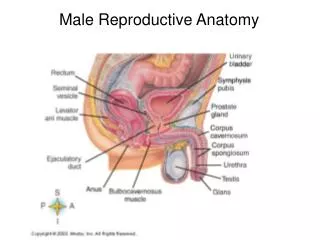
B. The external genitalia • The external genitalia arise from the same structures in both sexes –
At 8 weeks the external structures appear • include a genital tubercle, the genital folds, urethral folds and a • Genital tubercle gives rise to the clitoris and the urethral groove persists as the vestibule. • The unfused utrethral folds become the labia minora.
Development review • two products of the developing testes are needed for normal male development. • In the female, absence of androgens permits the external genitalia to remain feminine
C. Hormonal imbalance results in variation: • Any interference with the normal pattern of sex hormone production in the embryo • If testes do not produce testosterone – XY • If testes do not produce AMH, both female and male duct systems form, but the external genitalia are male • If XX is exposed to testosterone –
Psuedohermaphroditism • Androgen insensitivity syndrome (AIS) • If the testes form only partially and the production of androgen and Mullerian inhibitor is incomplete, there will be only partial masculinization of the external genitalia as well as incomplete inhibition of development of the vagina, uterus and Fallopian tubes. • If the testes form normally but are enzymatically incapable of completion of testosterone metabolism, there will be failure of complete masculinization of the external structures. However, the inhibition of Mullerian ducts will be normal because of normal production of Mullerian inhibitor, and therefore the upper vagina, uterus and Fallopian tubes may be absent. • Congenital adrenal hyperplasia • The androgen from the adrenal causes masculinization of the external genitalia, but since there is no Mullerian inhibitor, a normal uterus and vagina will develop
Psuedohermaphrodites – individuals with accessory reproductive structures that do not “match” their gonadsTrue hermaphrodites – rare, possess both ovaries and testicular tissue
CAH AIS
Genetic imbalance due to nondisjunction Sex-chromosome differences more frequent than autosomal abnormalities: • X inactivation • Y contains few functional genes • XO – Turner syndrome (female) • YO • XXX – Trisomy X (female) • XXXX • XXY – Klinefelter syndrome (male) • XYY – Double Y (male)
Klinefelter Syndrome Turner Syndrome
Production of haploid gametes is essential for sexual reproduction
III. Oogenesis A. Differentiation of the ovum • Forms a gamete that contains all the factors needed to initiate and maintain metabolism and development (as compared to simple sperm) • Haploid nucleus • Cytoplasmic enzymes • mRNAs • organelles • metabolic substrates • gametes develop from primordial germ cells that migrate to the somatic gonadal precursors (genital ridges in mammals) same for drosophila, xenopus & zebrafish • once the gonad is assembled, germ cells begin active proliferation and become gonial cells.
B. Follicle formation • In all species except C. elegans, gonadal somatic cells envelop female germ cells to create a follicle • Primordial Germ cells destined to become oocytes have already entered meiosis at the time of follicle formation and within the follicle they reach the diplotene stage of PI – here they halt meiosis for a few days to many years, depending on the species • Mechanisms of oogenesis differ among taxa because reproduction patterns also vary
Synchronous development leads to "cohorts" of eggs which are in the same stage of development, which reach maturity at the same time, and which are spent all at once. Hence, synchroneous gonad development is a cyclical affair: The gonad grows in size and mass during the ripening of the eggs until a distinct spawning event occurs which sets the gonad back to the initial conditions of the cycle In the gonads of species with asynchronous development we will not find "cohorts" of eggs being in the same stage of development, but there are eggs of any develop-mental stage at any time. These species show no distinct spawning times, but more or less continuous spawning all year round.
1). In Drosophila, asymmetric cell division gives rise to the oocyte • Cystoblast undergoes 4 mitotic divisions to give rise to a cyst of 16 germline cells http://berglab.gs.washington.edu/movies/cy2xGFPmoe.qt
2). Oogenesis in Vertebrates • Cyst phase is confined to early ovarian development and cells in an individual cyst do not differentiate as nurse cells
it is during oogenesis that the animal-vegetal axis of the egg is specified. as a result of differential intracellular transport, the amount of yolk steadily increases in the vegetal hemisphere Egg asymmetry
Oogenesis in Xenopus Iaevis • Oogonia are found as nests of 16 pearl shaped cells that develop synchronously
Growth of oocytes in the frog. During the first 3 years of life, three cohorts of oocytes are produced. The drawings follow the growth of the first-generation oocytes.
The large cell in the picture above is either the product of the first or second meiotic division that lives.
Follicular growth stage & antrum formation • Ooctye undergoes • increase in # of follicular granulosa cells • Cavity forms (antrum) which fills with proteins, hormones & additional molecules • Follicle cells secrete growth & differentiation factors
The ovarian follicle of mammals. (A) Maturation of the ovarian follicle. When mature, it is often called a Graafian follicle. (B) Scanning electron micrograph of a mature follicle in the rat.