Fisiologia Renal
460 likes | 734 Vues
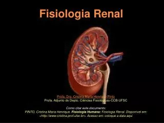
Fisiologia Renal. Mecanismos tubulares renais: segunda parte. Profa. Dra. Cristina Maria Henrique Pinto Profa. Adjunto do Depto. Ciências Fisiológicas-CCB-UFSC Como citar este documento:

Fisiologia Renal
E N D
Presentation Transcript
Fisiologia Renal Mecanismos tubulares renais: segunda parte Profa. Dra. Cristina Maria Henrique Pinto Profa. Adjunto do Depto. Ciências Fisiológicas-CCB-UFSC Como citar este documento: PINTO, Cristina Maria Henrique. Fisiologia Humana: Fisiologia Renal. Disponível em: <http://www.cristina.prof.ufsc.br>.Acesso em: coloque a data aqui
Bibliografia básica recomendada sobre Fisiologia Humana Livros-textos: “Berne & Levy: Fisiologia” Koeppen & Stanton, 2009, 6ª Ed. (Ed. Elsevier) “Tratado de Fisiologia Médica” Guyton & Hall, 2006, 11ª Ed. (Ed. Elsevier) “Fisiologia” Aires, M. M. 2008, 3ª Ed. (Ed. Guanabara Koogan) “Fisiologia” Costanzo, 2007, 3ª Ed. (Ed. Elsevier) “Berne & Levy: Fundamentos de Fisiologia”, Levy et al, 2006, 4ª Ed. (Ed. Elsevier) “Fundamentos de Fisiologia Médica” Johnson, 2003 (Ed. Guanabara Koogan) “Fisiologia: texto e atlas” Silbernagl e Despopoulos, 2003 (Ed. Artmed)
AS FIGURAS AQUI UTILIZADAS FORAM RETIRADAS DE DIVERSOS WEBSITES E, QUANDO POSSÍVEL, SÃO SEGUIDAS PELO ENDEREÇO NA INTERNET. PARA CONSULTA A TEXTOS E OUTROS RECURSOS ONLINE, VISITE OS SITES RECOMENDADOS EM “SITES DIDÁTICOS” PRESERVE O DIREITO AUTORAL CITANDO A FONTE.
3º seminário: Mecanismos tubulares renais 2 Alça de Henle e TCDinicial Alça de Henle: Reabsorção de 10% do volume filtrado: 10% da água (descendente) 20% do NaCl (ascendente) 20% do Ca++, K+ e Mg++
Reabsorção de Água e NaCl na Alça de Henle extraído, enquanto disponível, de: http://sprojects.mmi.mcgill.ca/nephrology/presentation/index.htm
Reabsorção de Água e NaCl na Alça de Henle Porção fina ou descendente AQP 1 extraído, enquanto disponível, de: http://sprojects.mmi.mcgill.ca/nephrology/presentation/index.htm
Distribuição dos subtipos de aquaporinas no néfron Diagrammatic representation of the localization of different aquaporins in the nephron and collecting duct system. AQP1 (blue) is present in the proximal tubule and descending thin limb. AQP2 (green) is abundant in the apical and subapical part of collecting duct principal cells, whereas AQP3 (red) and AQP4 (purple) are both present in the basolateral plasma membrane of collecting duct principal cells. AQP7 (orange) is confined to the apical brush border of straight proximal tubules. ADH, antidiuretic hormone. Nielsen et al., 2002 Physiol. Rev. 82: 205-244 Caso interesse saber um pouco mais sobre as aquaporinas, veja Verkman, 2005: “More than just water channels: unexpected cellular roles of aquaporins.” http://www.ncbi.nlm.nih.gov/entrez/query.fcgi?cmd=Retrieve&db=pubmed&dopt=Abstract&list_uids=16079275&query_hl=3&itool=pubmed_DocSum
Reabsorção de Água e NaCl na Alça de Henle AQP 1 extraído, enquanto disponível, de: http://sprojects.mmi.mcgill.ca/nephrology/presentation/index.htm
Reabsorção de Água e NaCl na Alça de Henle Porção ascendente AQP 1 extraído, enquanto disponível, de: http://sprojects.mmi.mcgill.ca/nephrology/presentation/index.htm
Reabsorção de Água e NaCl na Alça de Henle Porção ascendente FINA AQP 1 extraído, enquanto disponível, de: http://sprojects.mmi.mcgill.ca/nephrology/presentation/index.htm
Reabsorção de Água e NaCl na Alça de Henle ESPESSA Porção ascendente FINA AQP 1 extraído, enquanto disponível, de: http://sprojects.mmi.mcgill.ca/nephrology/presentation/index.htm
Reabsorção na Alça de Henle água (fina ou descendente)e NaCl (ascendente fina e espessa) Região medular renal AQP 1 extraído, enquanto disponível, de: http://sprojects.mmi.mcgill.ca/nephrology/presentation/index.htm
veja texto com explicações em: http://www2.kumc.edu/ki/physiology/index.htm
veja texto com explicações em: http://www2.kumc.edu/ki/physiology/index.htm
veja texto com explicações em: http://www2.kumc.edu/ki/physiology/index.htm
veja texto com explicações em: http://www2.kumc.edu/ki/physiology/index.htm
veja texto com explicações em: http://www2.kumc.edu/ki/physiology/index.htm
veja texto com explicações em: http://www2.kumc.edu/ki/physiology/index.htm
veja texto com explicações em: http://www2.kumc.edu/ki/physiology/index.htm
Reabsorção de NaCl na Alça de Henle (porção espessa ascendente)
http://www.alternex.com.br/~rfaria/ AHae e TCDi Fig. 17. Cellular mechanism of sodium, potassium, and anion transport in thick ascending limbs of Henle's loop. Arrows indicate net fluxes of solutes. The names of the currently cloned transporters are mentioned into rectangular boxes. Féraille and Doucet, 2001
http://www.alternex.com.br/~rfaria/ AHae e TCDi Reabsorção de outros íons Via paracelular Fig. 17. Cellular mechanism of sodium, potassium, and anion transport in thick ascending limbs of Henle's loop. Arrows indicate net fluxes of solutes. The names of the currently cloned transporters are mentioned into rectangular boxes. Féraille and Doucet, 2001 Féraille and Doucet, 2001
Reabsorção de NaCl na Alça de Henle (porção espessa ascendente) K+ extraído, enquanto disponível, de: http://sprojects.mmi.mcgill.ca/nephrology/presentation/index.htm
Inibição da reabsorção de NaCl na AH pelo Furosemide (“diurético de alça”) Alça de Henle (porção espessa ascendente) Furosemide extraído, enquanto disponível, de: http://sprojects.mmi.mcgill.ca/nephrology/presentation/index.htm
Reabsorção de Bicarbonato (HCO3-) no túbulo contorcido proximal 80-90% Na+ 2 c.a. capilar extraído, enquanto disponível, de: http://sprojects.mmi.mcgill.ca/nephrology/presentation/index.htm
Reabsorção de Bicarbonato (HCO3-) na Alça de Henle espessa 10-15% Na+ 2 3 c.a. AH não possui ca nos vilos capilar extraído, enquanto disponível, de: http://sprojects.mmi.mcgill.ca/nephrology/presentation/index.htm
Manipulação renal de Cálcio (Ca++) 11% do Cálcio plasmático são filtrados TCP reabsorve 70% do que foi filtrado (20% dos 55% de cálcio livres no plasma) Demais 45%: ligados às ptn dependente da reabsorção de Na+ e águano TCP (paracelular) extraído, enquanto disponível, de: http://sprojects.mmi.mcgill.ca/nephrology/presentation/index.htm
Manipulação renal de Cálcio (Ca++) TCP reabsorve 60% Dos 30%: 20% reabs. na AHae - TCDin Furosemide Excreção de Ca++ Dependente da reabsorção de Na+/K+/2Cl- (paracelular) extraído, enquanto disponível, de: http://sprojects.mmi.mcgill.ca/nephrology/presentation/index.htm
Manipulação renal de Cálcio (Ca++) Demais 10%: 8% podem ser reabsorvidos no TCD final na presença de PTH extraído, enquanto disponível, de: http://sprojects.mmi.mcgill.ca/nephrology/presentation/index.htm
Mechanism of Ca2+ homeostasis. Figure 1 An important element of total body Ca2+ homeostasis is the sensing of blood Ca2+ levels by the Ca2+-sensing receptor (CaR) in parathyroid cells. Following a decrease in blood Ca2+ levels, this receptor triggers the release of the parathyroid hormone (PTH). This in turn leads to Ca2+ release from bone and enhanced 1,25-vitamin D (1,25-VitD) production from 25-vitamin D (25-VitD) in the proximal tubule of the kidney. 1,25-Vitamin D increases the expression of the epithelial Ca2+ channels TRPV6 and, together with PTH, TRPV5. TRPV6 transports Ca2+ into the body from the small intestine across brush border membranes, whereas TRPV5 enhances Ca2+ reabsorption in the distal convoluted tubule in the kidney. extraído, enquanto disponível, de: http://arjournals.annualreviews.org/doi/full/10.1146/annurev.physiol.69.031905.161003
Mechanism of Ca2+ homeostasis. Figure 1 An important element of total body Ca2+ homeostasis is the sensing of blood Ca2+ levels by the Ca2+-sensing receptor (CaR) in parathyroid cells. Following a decrease in blood Ca2+ levels, this receptor triggers the release of the parathyroid hormone (PTH). This in turn leads to Ca2+ release from bone and enhanced 1,25-vitamin D (1,25-VitD) production from 25-vitamin D (25-VitD) in the proximal tubule of the kidney. 1,25-Vitamin D increases the expression of the epithelial Ca2+ channels TRPV6 and, together with PTH, TRPV5. TRPV6 transports Ca2+ into the body from the small intestine across brush border membranes, whereas TRPV5 enhances Ca2+ reabsorption in the distal convoluted tubule in the kidney. extraído, enquanto disponível, de: http://arjournals.annualreviews.org/doi/full/10.1146/annurev.physiol.69.031905.161003
Mechanism of Ca2+ homeostasis. Figure 1 An important element of total body Ca2+ homeostasis is the sensing of blood Ca2+ levels by the Ca2+-sensing receptor (CaR) in parathyroid cells. Following a decrease in blood Ca2+ levels, this receptor triggers the release of the parathyroid hormone (PTH). This in turn leads to Ca2+ release from bone and enhanced 1,25-vitamin D (1,25-VitD) production from 25-vitamin D (25-VitD) in the proximal tubule of the kidney. 1,25-Vitamin D increases the expression of the epithelial Ca2+ channels TRPV6 and, together with PTH, TRPV5. TRPV6 transports Ca2+ into the body from the small intestine across brush border membranes, whereas TRPV5 enhances Ca2+ reabsorption in the distal convoluted tubule in the kidney. extraído, enquanto disponível, de: http://arjournals.annualreviews.org/doi/full/10.1146/annurev.physiol.69.031905.161003
Mechanism of Ca2+ homeostasis. Figure 1 An important element of total body Ca2+ homeostasis is the sensing of blood Ca2+ levels by the Ca2+-sensing receptor (CaR) in parathyroid cells. Following a decrease in blood Ca2+ levels, this receptor triggers the release of the parathyroid hormone (PTH). This in turn leads to Ca2+ release from bone and enhanced 1,25-vitamin D (1,25-VitD) production from 25-vitamin D (25-VitD) in the proximal tubule of the kidney. 1,25-Vitamin D increases the expression of the epithelial Ca2+ channels TRPV6 and, together with PTH, TRPV5. TRPV6 transports Ca2+ into the body from the small intestine across brush border membranes, whereas TRPV5 enhances Ca2+ reabsorption in the distal convoluted tubule in the kidney. extraído, enquanto disponível, de: http://arjournals.annualreviews.org/doi/full/10.1146/annurev.physiol.69.031905.161003
Mechanism of epithelial Ca2+ absorption in the intestine and kidney. Figure 2. In theintestine, the TRPV6 epithelial Ca2+ channel, which is expressed in thebrushbordermembrane, mediatesthefirststep in transepithelial Ca2+ absorption. Onceinsidetheepithelialcells, Ca2+ binds to calbindin D9K. Calbindin D9K is thought to play a central role in transportingintracellular Ca2+ to thebasolateralmembranewithoutincreasingfree-Ca2+ concentration. Atthebasolateralmembrane Ca2+ is releasedintothebloodthroughthe Ca2+-ATPase PMCA1b andpossiblythe Na+/Ca2+ exchanger NCX1 (SLC8A1). Ca2+ reabsorption in the distal convolutedtubuleofthekidneyproceeds in a similar fashion butwiththefollowingvariations: (a) Ca2+ entryatthe luminal side is mediatedpredominantlybytheepithelial Ca2+ channel TRPV5. (b) Ca2+ is shuttled to thebasolateralmembrane via bothcalbindin D9K andcalbindin D28K. (c) Ca2+ exitproceeds via both PMCA1b Ca2+-ATPaseandthe NCX1 (SLC8A1) Na+/Ca2+ exchanger. Underhigh luminal Ca2+ conditions, Ca2+ is absorbed, via theparacellularroute, throughthetightjunctiondownthetransepithelial Ca2+ gradient. extraído, enquanto disponível, de: http://arjournals.annualreviews.org/doi/full/10.1146/annurev.physiol.69.031905.161003
Manipulação renal de Cálcio (Ca++) extraído, enquanto disponível, de: http://sprojects.mmi.mcgill.ca/nephrology/presentation/index.htm
Manipulação renal de Cálcio (Ca++) extraído, enquanto disponível, de: http://sprojects.mmi.mcgill.ca/nephrology/presentation/index.htm
Manipulação renal de Cálcio (Ca++) Reabsorção de até 8% do que foi filtrado. Excreção de, no mínimo, 2% Aumenta a reabsorção renal de cálcio (diminui a excreção) extraído, enquanto disponível, de: http://sprojects.mmi.mcgill.ca/nephrology/presentation/index.htm
Manipulação renal de Cálcio (Ca++) Aumenta a absorção intestinal de cálcio Aumenta a reabsorção renal de cálcio (diminui a excreção) extraído, enquanto disponível, de: http://sprojects.mmi.mcgill.ca/nephrology/presentation/index.htm
Reabsorção de Cálcio (Ca++) dependente de PTH (TCD) 3 Na+ 3 Na+ Ca++ Ca++ Ca++ extraído, enquanto disponível, de: http://sprojects.mmi.mcgill.ca/nephrology/presentation/index.htm
Reabsorção de Cálcio (Ca++) dependente de PTH (TCD) PTH e Calcitriol PTH e Calcitriol + 3 Na+ 3 Na+ Ca++ + Ca++ Ca++ extraído, enquanto disponível, de: http://sprojects.mmi.mcgill.ca/nephrology/presentation/index.htm
Reabsorção de NaCl pelo TCD 3 a 5% do que foi filtrado NaCl extraído, enquanto disponível, de: http://sprojects.mmi.mcgill.ca/nephrology/presentation/index.htm
Reabsorção de NaCl pelo TCD Diuréticos Tiazídicos (Hidroclorotiazida) 2 3 Cl- Cl-
Reabsorção de Cálcio (Ca++) dependente de PTH (TCD) Cl- Cl- Diuréticos Tiazídicos inibem a reabsorção de NaCl (3-5%) Aumentam a reabsorção de Cálcio Cl- 3 Na+ 3 Na+ Ca++ Ca++ Ca++ extraído, enquanto disponível, de: http://sprojects.mmi.mcgill.ca/nephrology/presentation/index.htm
Manipulação renal de Cálcio (Ca++) Demais 10%: 8% podem ser reabsorvidos no TCD final na presença de PTH extraído, enquanto disponível, de: http://sprojects.mmi.mcgill.ca/nephrology/presentation/index.htm
FISIOLOGIA RENAL continua em: Aspectos integrados das funções renais Veja também: Introdução ao estudo da Fisiologia Renal Mecanismos tubulares I Veja outros recursos sobre Fisiologia Renal nos links abaixo: Material didático de Renal, extraído da Internet (apresentações em ppt, capítulos de livros em pdf, animações online, etc) Sugestões de sites didáticos de Fisiologia Renal