Advanced Focused Ion Beam: Cobalt Alloy Microstructure Analysis
100 likes | 175 Vues
Explore magnetic cobalt alloy microstructure using Focused Ion Beam technique with detailed specimen preparation steps including sectioning and thinning using Gallium ion beam.

Advanced Focused Ion Beam: Cobalt Alloy Microstructure Analysis
E N D
Presentation Transcript
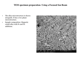
TEM specimen preparation- Using a Focused Ion Beam • The alloy microstructure is shown alongside. It has a two-phase microstructure. • Sample composition: Magnetic cobalt alloy with Fe and Ni additions.
After selecting the area to investigate, a micron thick strip of Platinum is electro-deposited to protect the area beneath from being contaminated by the Gallium ions. • The electron beam is maintained at 5 kV and a current of 6.3nA.
The selection is made to mill away the specimen on both sides of the Pt strip using the Gallium ion beam maintained at 30 kV and 7 nA. ION BEAM VIEW ELECTRON BEAM VIEW
The other side of the Pt bar suffers the same fate. • Further thinning of the specimen on either side of the Pt strip is carried out at a reduced ion current of 3 nA. (see next page)
FURTHER THINNING ION VIEW ION VIEW ELECTRON VIEW ELECTRON VIEW
The sample is tilted to 7º (from horizontal) and areas for the release cuts are marked, and the approximate z-depth (on the order of the thickness of the thinned slab) is entered and the selected areas are cut… The ion current is decreased further to 0.3 nA.
Pt deposition at the probe tip binds it tightly to the strip
The final release cuts are now made to free the specimen from the underlying bulk. These cuts are made at very low current of 10 pA. • The final polishing (see next slide) is made to thin the specimen even further with an ion beam current of 100 pA.