Chapter 3- Cell Structure
Chapter 3- Cell Structure. I. Looking at cells. A. Microscopes Reveal cell structure 1. Most cells are too small to see with the naked eye, a typical human body cell is many times smaller than grain sand.

Chapter 3- Cell Structure
E N D
Presentation Transcript
I. Looking at cells A. Microscopes Reveal cell structure 1. Most cells are too small to see with the naked eye, a typical human body cell is many times smaller than grain sand. 2. Robert Hooke first saw the cell and explained what he saw as, “a lot of little boxes.”
2. Anton van Leeuwenhoek discovered “animalcules,” today we know them as single celled organisms
B. Measuring the sizes of cell structure 1. Measurements taken by scientists are expressed in metric units. 2. Scientists throughout the world use the metric system.
3. The official name- International System of Measurement- SI. 4. SI is a decimal system, so all relationships are based on powers of ten. 5. Look and KNOW table 3.1
C. Characteristics of microscopes 1. In a light microscope, light passes through one or more lenses to produce an enlarged image of a specimen.
2. An electron microscope forms an image of a specimen using a beam of electrons rather than light.
3. Magnification is the ability to make an image appear larger than its actual size. 4. Resolution is a measure of the clarity of an image. 5. Both magnification and resolution is needed to view microscopic organisms.
D. Microscopes have different uses and limitations 1. Light microscopes that use two lenses are called compound light microscopes. 2. Compound microscopes can have a total magnification up to 2,000X
3. Electron microscopes can magnify an image up to 200,000X and they can be used to study even the smallest structures inside cells or on the cell surface. 4. In transmission electron microscopes the electron beam is directed at a very thin slice of a specimen stained with metal ions.
5. Scanning electron microscopes the electron beam is focus on a specimen coated with a very thing layer of metal. 6. The scanning tunneling microscope uses a needle-like probe to measure differences in voltage caused by electrons that leak, or tunnel from the surface of the objects being viewed.
II. Cell Features A. Schwann and Virchow form the cell theory which has three parts: 1.All living things are made up of one or more cells. 2.Cells are the basic units of structure and function in organisms. 3.All cells arise from existing cells.
B. Cells must be small 1. They are small but they contain everything that is needed.
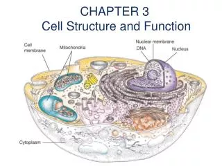
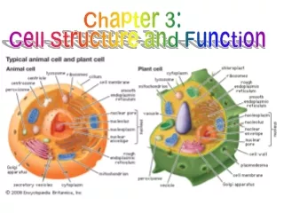
C. Common features of cell 1. Cells share common structure features, including an outer boundary called the cell membrane. The cell membrane encloses the cell and separates the cell. 2. Cytoplasm is the jelly like substance of the cell.
3. Inside the cytoplasm are many structures, often suspended in a system of microscopic fibers called cytoskeleton.
B. Prokaryotes do not contain internal compartments 1.A prokaryote is a single-celled organism that lacks a nucleus and other internal compartments. 2.Modern prokaryotes are called bacteria.
3. The cytoplasm of a bacterial cell includes everything inside the cell membrane. 4. Bacterial cells have a cell wall surrounding the cell membrane that provides structure and support. 5. Many bacteria have flagella, which are long, threadlike structures that allow the bacteria to move.
D. Eukaryotic cells are organized 1. The first cells with internal compartments appeared 1.5 billion years ago. 2. A eukaryote is an organism whose cells have a nucleus.
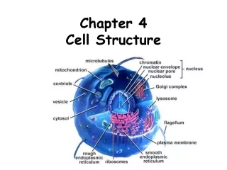
3. The nucleus is an internal compartment that houses the cell’s DNA. 4. An organelle is a structure that carries out specific activities in the cell. 5. Short hair like structures is called cilia. 6. See figure 3.8 to see an Eukaryotic cell.
F. The structure and Function of cell membranes are closely related. 1. The selective permeability of the cell membrane is caused mainly by a phospholipid. 2. In a cell membrane, the phospholipids are arranged in a double layer called a lipid belayed. 3.See figure 3.9
4. Various proteins are located in the lipid bilayer of a cell membrane. 5. Cell membranes contain different types of proteins, each plays a vital role in the life of a cell.
III. Cell Organelles A. The Nucleus Directs Cell Activities and Stores DNA 1.Most functions of a Eukaryotic cell area controlled by the cell’s nucleus. 2.A double membrane called the nuclear envelope, also called the nuclear membrane, surrounds the nucleus. 3.The hereditary information of a Eukaryotic cell is coded in the cell’s DNA, which is stored in the nucleus.
B. An internal membrane system processes proteins 1.Unlike prokaryotic cells, Eukaryotic cells have a system of internal membranes that play an essential role in the processing of proteins.
C. Production of proteins 1.The endoplasmic reticulum (ER) moves proteins and other substances through the cell. It acts like a highway. 2.There is rough and smooth ER. Rough ER has ribosomes attached to it and smooth ER does not.
3. As each protein is made, it crosses the ER membrane and enters the ER. The portion of the ER that contains the completed protein then pinches off to form a vesicle. 4. A vesicle is a small, membrane- bound sac that transports substances in cell.
D. Packaging and distributing of proteins 1. The Golgi apparatus is a set of flattened, membrane-bound sacs that serves as the packaging and distributing center of the cell. It acts like the UPS of the cell. 2. The cell also has lysosomes. They are vesicles, which contain digestive enzymes. They eat the garbage of the cells. “Suicide bags.”
E. Steps of the Golgi 1.Ribosomes make proteins and hop on the ER for a ride. 2.The ER takes them to the Golgi. 3.The Golgi repackages the proteins into what the cell needs.
4. Golgi spits out the new vessels and they go where they are needed. 5. Lysosomes go around and eat up the old or damaged vessels.
F. Mitochondria Produces ATP 1.Nearly all Eukaryotic cells contain many mitochondria. 2.Mitochondrion makes ATP (energy) for the cell. “Powerhouse of the cell.”
G. Plant cells contain structures that animal cells lack 1.The cell membrane of a plant cell is surrounded by a cell wall. (Animal cells DO NOT have a cell wall) 2.The cell wall helps to support and maintain the cell’s shape, protects the cell from damage. 3. It DOES NOT prevent in things in our out of the cell this is the cell membranes job.
4. Plant cells contain one or more chloroplasts. 5. Chloroplasts are organelles that use light energy to make food. 6. Chloroplasts make chlorophyll. 7. Much of the plant cell’ s volume is taken up by a large; membrane bound space called the central vacuole.