Download
1 / 54
640 likes | 1.18k Vues
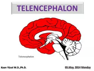
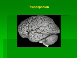
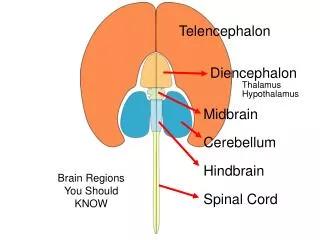
Telencephalon. Diencephalon Thalamus Hypothalamus. Midbrain. Cerebellum. Hindbrain. Brain Regions You Should KNOW. Spinal Cord. D o r s a l – S e n s o r y. Thalamus. V e n t r a l – M o t o r. Hypothalamus. Visual Pathways from Retina to CNS.

E N D
Telencephalon Diencephalon Thalamus Hypothalamus Midbrain Cerebellum Hindbrain Brain Regions You Should KNOW Spinal Cord
D o r s a l – S e n s o r y Thalamus V e n t r a l – M o t o r Hypothalamus
Visual Pathways from Retina to CNS • Know the basic organization of visual system pathways, from retina to primary visual cortex and then continuing to other visual/cortical areas for processing of color, movement, and complex shapes (e.g., human faces). • Know the function of midbrain and hypothalamic pathways. • Know the differences in visual system organization between animals with eyes on the sides of the head (monocular depth perception) vs. front of the face (binocular depth perception). • Why do only the nasal retinas cross in humans? In other words, what would we see if these pathways did NOT cross? How does this relate to retinal disparity and depth/space perception?
Somewhere in Cortex But don’t forget! We’ll have to do this 3 times, and maybe a 4th! Color Shape Movement The Retina (ganglion cell receptive fields)
Somewhere in Cortex But don’t forget! We’ll have to do this 3 times, and maybe a 4th! Color Shape Movement The Retina (ganglion cell receptive fields)
Primary Visual Cortex Optic Nerve LGN (thalamus) Midbrain SCN (hypothalamus) Dorsal Ventral • Where does light (‘dots of contrast’) information go? • Suprachiasmatic Nucleus (SCN, Biological Clock) • What vs. Where (Cortex vs. Midbrain) • Blind Sight (Midbrain) vs. Conscious Sight (Cortex)
Prey animal:eyes on side of the head - each eye gets a different image Normally, visual pathways ‘cross’ – the left half of the visual field is processed in the right half of the brain, and vice versa. WHY? Well, with thanks to the clever Cajal, let’s have a look at what would happen if the visual pathways did NOT cross… An incoherent image would be produced in the brain
Human OR Predator:Two (slightly different) Pictures of the Same ThingYour ocular muscles insure that the image in each eye is split equally across nasal and temporal retinas Color Shape Movement The visual thalamus has 6 layers, 3 for each eye.
What happens when you cross your eyes?Now the image is projected onto only one half of the retina.Two important things happen… d = optic nerve c = optic chiasm g = thalamus Rv = right visual ctx
The purpose of the two images is to generate ‘binocular disparity’, which is greater when objects are close to us. The visual system estimates the depth of an object based on binocular disparity.
Movement 3 ‘channels’ leave the Retina Color Shape Movement Color Each channel is destined for its own cortical processing area It all begins here… Shape Face Processing
VISUAL PROCESSING IN CORTEX • Know the basic organization and function of primary visual cortex (the hypercolumn!). • As we move from retina to cortex, the response properties of cells in the visual system become increasingly complex. What synaptic mechanisms are used to produce cells that could respond to complex shapes (e.g., Geons) or movement? • Perception of Motion (why the retina can’t know whether something is moving or not).
retina Eye-specific Layers thalamus Eye-specific Columns cortex A Surprise: The First Elements of Shape Are Little Bars
retina retina thalamus Input Cells thalamus cortex V1 Cortical Cell cortex
retina retina thalamus Input Cells thalamus cortex V1 Cortical Cell cortex
Primary Visual Cortex is a VAST ARRAY of HYPERCOLUMNS(this example shows three hypercolumns) Occular Dominance Columns Orientation Columns 3 2 1 Each column responds to a ‘bar’ of contrast with a specific orientation (Orientation Columns – specified by response). Orientation Columns are organized into Left and Right eye groups (Occular Dominance Columns – specified by input).
What does this mean? Occular Dominance Columns Orientation Columns 3 2 1 A single Hypercolumn corresponds to ONE point in visual space. Everything that you could possibly see in that ONE point is already represented in the corresponding Hypercolumn. In other words, everything you can see is already represented in Primary Visual Cortex. If it isn’t there, you can’t see it.
What happens when I show you an ‘X’ in your LEFT visual field? Left nasal retina Right temporal retina L R L R Right Primary Visual Cortex
What does the visual system do with all these little bars?
3 ‘dot’ detecting cells converge on a single ‘bar’ detecting cell in primary visual cortex (V1) retina thalamus Input Cells cortex V1 Cortical Cell Somewhere downstream, these 4 ‘bar’ detecting responses converge on a single ‘square’ detecting cell
Example of responses of a V2 cortical neuron to visual stimuli Hedge and Van Essen, 2000
3 ‘square’ detecting cells converge on a single ‘cube’ detecting cell located in a tertiary processing area – etc., etc., … Geons hypothesized to be ‘3D Shape Primaries’
Movement 3 ‘channels’ leave the Retina Color Shape Color Movement Each channel is destined for its own cortical processing area It all begins here with little dots of contrast… Shape Face Processing
Many:1 patterns of connections can be used to make a ‘moving bar’ detecting cell Delay lines. . . I see a vertical bar of contrast moving left to right!
Many:1 patterns of connections can be used to make a ‘moving bar’ detecting cell Delay lines. . . I see a vertical bar of contrast moving left to right!
What happens if the bar is moving too fast? Delay lines. . . Wha!!??
Star Moves Right Why Motion Can’t Be ‘Sensed’ Eye Moves Right The Pattern of Image Movement on the Retina is the Same!
For More On The Visual Thalamus and visual cortex see ch. 3 of your book… • PAGE 62: The Lateral Geniculate Nucleus • 6 Layers (3 from each eye – shape, color, movement!) • Top 4 layers (parvocellular layers) are shape and color • Bottom 2 layers (magnocellular layers) are movement • PAGE 64: Striate Cortex (aka ‘Primary Visual Ccortex’, aka ‘V1’) • Cortical magnification (most of your visual cortex is being used to process information from your fovea – this is very similar to somatosensory cortex, where the hands and face occupy a disproportionate amount of cortex – remember the homunculus!) • PAGE 68: Receptive fields in Striate Cortex • Very similar to what I’ve talked about in class – cells respond to ‘bars’ of various orientations. • PAGE 72: Columns and Hypercolumns • Should match up with the lecture material
What can visual cortex teach us about the nature/nurture debate? • Review ‘Nativism and Empiricism’ (p. 7 and 8 in your book). • What role is played by experience in the development of ocular dominance, orientation columns, and the visual system in general? How does this information help us understand ‘critical periods’ during development? What are the implications of these data for thinking about the roles of nature vs. nurture in human development?
Effects of Experience on Ocular Dominance Columns Nativism vs. Empricism
Put Tracer in One Eye ‘Seeing’ Ocular Dominance Columns
Effects of Experience on Ocular Dominance Columns Nativism vs. Empricism
Effects of Experience on Occular Dominance Columns Normal Development Raise Animal In The Dark Adult
Sorry Empiricists! Raise Animal In The Dark Adult
No Depth Perception Possible What Happens If Sight Returns in Adulthood? Raise Animal In The Dark Adult
What is ‘teaching’? Are short attention spans in youngsters ‘abnormal’? Remember empiricism? Critical Periods and Puberty
Critical Period Lifespan 100 80 60 Chemical Element No. 40 20 R. Buckminster Fuller 0 0 1K 2K 3K 4K 5K Recorded History • Key Points from this graph: • Nature assumes that the environment will change slowly, but in an unpredictable way – hence, ‘Critical Periods’ during early development. • Critical periods of development worked just fine several thousand years ago. • However, our cultural environment is now changing at an ever-increasing rate, violating Nature’s assumption of slow change. We need to keep learning NEW THINGS throughout our lifespan – new emphasis on ‘lifelong learning’.
Comments on Chapter 4: How does this relate to lecture material? This chapter starts off with a discussion of ‘Middle Vision’ that includes a bunch of Gestalt Illusions. No need to memorize the names of these illusions, but it is VERY important to understand the historical significance of Gestalt Psychology. This is where the distinction between ‘sensation’ and ‘perception’ originates. The only way to explain Gestalt illusions is to invoke some process beyond sensation. For example, in the classic ‘face/vase’ illusion (see Fig. 4.26) you will note that ONE retinal image gives rise to MULTIPLE perceptions. This is where we get the saying ‘the whole is greater than the sum of the parts’. Another way to say that is ‘the perception is greater than the sum of the sensation’. As we’ve said before in this class, you want to think of sensation as a passive, analytic process and perception as an active, synthetic process.
Comments on Chapter 4: How does this relate to lecture material? It is also worth thinking about the implications of Gestalt Psychology for Empiricism. The empiricists viewed the organism as a passive recipient of sensory information (an empty bucket, if you will). However, if an active, internal process of perception exists, notice that we cannot control exactly what goes in the ‘bucket’. The same sensation can be perceived differently by different individuals. Gestalt Psychology represented one of the first dents in the armor of Empiricism. The developmental studies on the visual cortex that we’ve discussed in class have likely cracked this armor open for good.
Comments on Chapter 4: How does this relate to lecture material? Notice the similarity between the ‘Pandemonium’ model (Figure 4.21) and the story I told in class (What does the visual system do with all these little bars?). Also note the discussion of geons on p. 109 of your book. As mentioned in lecture, complex shape processing occurs in the inferior temporal cortex. Damage to the inferior temporal cortex produces ‘prosopagnosia’ (face blindness). Beginning on p. 107 read the section ‘Object Recognition’ and continue reading through ‘Faces: An illustrative special case’.
Comments on Chapter 6: How does this relate to lecture material? This chapter begins with a large section on Monocular Cues to 3D space (time to get out your video games!). How can images on a 2D television or computer screen seem 3D? You’ll find the answers here. Pay special attention to ‘motion parallax’, which is perhaps the most powerful of the monocular cues for depth. Used all the time in video games, movies, and television. If an animal has its eyes on the sides of its head, all depth perception will be monocular, using these types of cues. We use these cues IN ADDITION to the more powerful and accurate Binocular Disparity mechanism discussed below. The next large section in Chapter 6 is on Binocular Vision. We’ve talked about this in lecture (that Cajal drawing in Slide 6). There is quite a bit of fancy terminology in the book, but bear in mind that all we are talking about is how the visual system uses DIFFERENCES between the two retinal images to COMPUTE depth perception (aka, stereopsis).