Download
1 / 45
450 likes | 517 Vues
Explore the arterial blood supply system, including pulmonary and systemic circulation, key arteries in the head, neck, upper limb, and abdomen, and clinical anatomy facts. Learn about important branching patterns and pulse points.

E N D
ARTERIAL BLOOD SUPPLY Dr. NabilKhouri
The Vascular system • The vascular system plays the critical role In: • Delivering nutrients • Clearing metabolic waste products from peripheral tissues • Maintaining systemic core temperature. Vascular flow is controlled by various processes, including: • Vessels anatomy & histology structure • Vascular tone, which is controlled by neuroendocrine hormones along with autonomic nervous system influence • End-organ metabolic requirements.
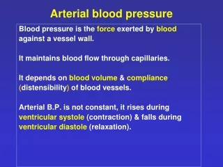
Two cardiovascular circulation • Pulmonary circulation • blood to and from the lungs • System circulation • blood to and from the rest of the body • Vessels carry the blood through these circuits • Arteries carry blood away from the heart • Veins carry blood to the heart • Capillaries permit exchange
Pulmonary Circulation • Pulmonary trunk branches • Right and left pulmonary arteries • Division into lobar arteries • 3 on right • 2 on left • Smaller and smaller arterioles, into capillaries surrounding alveoli • Gas exchange • Pulmonary system pressure is only 1/6 of systemic blood pressure • After gas exchange blood enters venules • Larger and larger into Superior and Inferior Pulmonary veins • Four Pulmonary Veins empty into left atrium
Systemic Circulation • Oxygenated blood to body • Leaves LV through Ascending Aorta • It has Only 2 branches: the left and right coronary arteries • Aortic Arch has three arteries branching from it: • Brachiocephalic trunk, has 2 branches: • Right common carotid a. • Right subclavian a. • Left common carotid a. • Left subclavian a.
Coronary arteries and sinuses Arteries of the Head and Neck
Internal carotid A. • Enters skull through carotid canal • Gives off: • Ophthalmic artery • Then divides into anterior and middle cerebral arteries together they supply 80% of cerebrum
Arteries of the Upper limb • Subclavian runs laterally onto 1st rib, under clavicle • Enters axilla as axillary artery • Sends branches • Continues as brachial artery in upper arm • Splits into radial & ulnar arteries • See hand supply
The axillary artery The axillary artery is separated into three parts by the pectoralis minor muscle, which crosses anteriorly to the vessel : • The first part is proximal to pectoralis minor; • The second part is posterior to pectoralis minor; • The third part is distal to pectoralis minor.
Generally, six branches arise from the axillary artery: • One branch, the superior thoracic artery, originates from the first part; • Two branches, the thoraco-acromial artery and the lateral thoracic artery, originate from the second part; • Three branches, the subscapular artery, the anterior circumflex humeral artery, and the posterior circumflex humeral artery, originate from the third part
Important clinical anatomy fact • In approximately 80% of patients, the deep and superficial palmar arches are connected and are referred to as complete. • This results in a Dual perfusion supply to the common and proper digital vessels. • This is an important attribute of hand vascular architecture, providing collateral blood flow in the event of vascular pathology affecting one of these palmar arches.
Descending Aorta • Anterior intercostals are branchs off the Internal thoracic*(A branch of subclavian) • Posterior intercostals are branchs off the Thoracic aorta
The Abdominal Aorta • At rest, ½ arterial blood is here! • Three single (Unpaired) midline branches supply the digestive tract • Celiac trunk • Superior mesenteric artery • Inferior mesenteric artery 1. 2. 3.
Celiac trunk: divides into 3 right away: left gastric, splenic & common hepatic 1. 2.
2. Superior mesenteric supplies most of intestines 3. Inferior mesenteric supplies distal half of large intestine 1. 2. 3.
Arteries to the abdomen • The Paired branches off the abdominal aorta supply adrenal glands, kidneys, gonads and abdominal body wall supply diaphragm supply adrenals to kidney 3.
Terminal branches of the abdominal Aorta • Abdominal aorta terminal branches are the two: the Rt and Lt Common iliacs • At L4; each will terminate by dividing into: • Internal iliacs to pelvic organs, perineum, buttocks, medial thighs • External iliacs: to rest of lower limbs
External iliac passes under inguinal ligament becoming Femoral artery • At back of knee femoral becomes popliteal artery, and branches Feel dorslis pedis & posterior tibial