Nucleic Acids
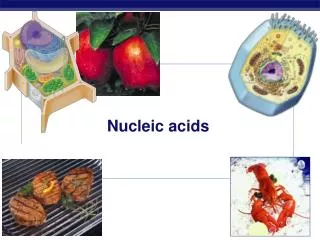
Nucleic Acids. IUG, Spring 2014 Dr. Tarek Zaida. Nucleic Acids. Nucleic acids are molecules that store information for cellular growth and reproduction There are two types of nucleic acids: - deoxyribonucleic acid (DNA) and ribonucleic acid (RNA)

Nucleic Acids
E N D
Presentation Transcript
Nucleic Acids IUG, Spring 2014 Dr. TarekZaida
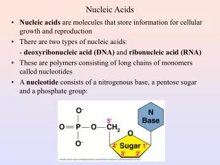
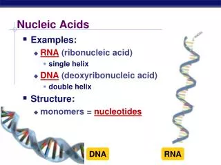
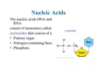
Nucleic Acids • Nucleic acids are molecules that store information for cellular growth and reproduction • There are two types of nucleic acids: - deoxyribonucleic acid (DNA) and ribonucleic acid (RNA) • These are polymers consisting of long chains of monomers callednucleotides • A nucleotide consists of a nitrogenous base, a pentose sugar and a phosphate group:
Nitrogen Bases • The nitrogen bases in nucleotides consist of two general types: - purines: adenine (A) and guanine (G) - pyrimidines: cytosine (C), thymine (T) and Uracil (U)
Pentose Sugars • There are two related pentose sugars: - RNA contains ribose - DNA contains deoxyribose • The sugars have their carbon atoms numbered with primes to distinguish them from the nitrogen bases
Nucleosides and Nucleotides • A nucleoside consists of a nitrogen base linked by a glycosidic bond to C1’ of a ribose or deoxyribose • Nucleosides are named by changing the nitrogen base ending to -osine for purines and –idine for pyrimidines • A nucleotide is a nucleoside that forms a phosphate ester with the C5’ OH group of ribose or deoxyribose • Nucleotides are named using the name of the nucleoside followed by 5’-monophosphate
AMP, ADP and ATP • Additional phosphate groups can be added to the nucleoside 5’-monophosphates to form diphosphates and triphosphates • ATP is the major energy source for cellular activity
Primary Structure of Nucleic Acids • The primary structure of a nucleic acid is the nucleotide sequence • The nucleotides in nucleic acids are joined by phosphodiester bonds • The 3’-OH group of the sugar in one nucleotide forms an ester bond to the phosphate group on the 5’-carbon of the sugar of the next nucleotide
Reading Primary Structure • A nucleic acid polymer has a free 5’-phosphate group at one end and a free 3’-OH group at the other end • The sequence is read from the free 5’-end using the letters of the bases • This example reads 5’—A—C—G—T—3’
Example of RNA Primary Structure • In RNA, A, C, G, and U are linked by 3’-5’ ester bonds between ribose and phosphate
Example of DNA Primary Structure • In DNA, A, C, G, and T are linked by 3’-5’ ester bonds between deoxyribose and phosphate
Secondary Structure: DNA Double Helix • In DNA there are two strands of nucleotides that wind together in a double helix - the strands run in opposite directions - the bases are arranged in step-like pairs - the base pairs are held together by hydrogen bonding • The pairing of the bases from the two strands is very specific • The complimentary base pairs are A-T and G-C - two hydrogen bonds form between A and T - three hydrogen bonds form between G and C • Each pair consists of a purine and a pyrimidine, so they are the same width, keeping the two strands at equal distances from each other.
DNA Replication • When a eukaryotic cell divides, the process is called mitosis - the cell splits into two identical daughter cells - the DNA must be replicated so that each daughter cell has a copy • DNA replication involves several processes: - first, the DNA must be unwound, separating the two strands - the single strands then act as templates for synthesis of the new strands, which are complimentary in sequence
- bases are added one at a time until two new DNA strands that exactly duplicate the original DNA are produced. • The process is called semi-conservative replication because one strand of each daughter DNA comes from the parent DNA and one strand is new • The energy for the synthesis comes from hydrolysis of phosphate groups as the phosphodiester bonds form between the bases
Ribonucleic Acid (RNA) • RNA is much more abundant than DNA • There are several important differences between RNA and DNA: - the pentose sugar in RNA is ribose, in DNA it’s deoxyribose. - in RNA, uracil replaces the base thymine (U pairs with A) - RNA is single stranded while DNA is double stranded - RNA molecules are much smaller than DNA molecules • There are three main types of RNA: - ribosomal (rRNA), messenger (mRNA) and transfer (tRNA)
Ribosomal RNA and Messenger RNA • Ribosomes are the sites of protein synthesis - they consist of ribosomal RNA(65%) and proteins (35%) - they have two subunits, a large one and a small one • Messenger RNA carries the genetic code to the ribosomes - they are strands of RNA that are complementary to the DNA of the gene for the protein to be synthesized
Transfer RNA • Transfer RNA translates the genetic code from the messenger RNA and brings specific amino acids to the ribosome for protein synthesis • Each amino acid is recognized by one or more specific tRNA • tRNA has a tertiary structure that is L-shaped - one end attaches to the amino acid and the other binds to the mRNA by a 3-base complimentary sequence
Protein Synthesis • The two main processes involved in protein synthesis are - the formation of mRNA from DNA (transcription) - the conversion by tRNA to protein at the ribosome (translation) • Transcription takes place in the nucleus, while translation takes place in the cytoplasm • Genetic information is transcribed to form mRNA much the same way it is replicated during cell division
Transcription • Several steps occur during transcription: - a section of DNA containing the gene unwinds - one strand of DNA is copied starting at the initiation point, which has the sequence TATAAA - an mRNA is synthesized using complementary base pairing with uracil (U) replacing thymine (T) - the newly formed mRNA moves out of the nucleus to ribosomes in the cytoplasm and the DNA re-winds
RNA Polymerase • During transcription, RNA polymerase moves along the DNA template in the 3’-5’direction to synthesize the corresponding mRNA • The mRNA is released at the termination point
Processing of mRNA • Genes in the DNA of eukaryotes contain exonsthat code for proteins along with introns that do not • Because the initial mRNA, called a pre-RNA, includes the noncodingintrons, it must be processed before it can be read by the tRNA • While the mRNA is still in the nucleus, the introns are removed from the pre-RNA • The exons that remain are joined to form the mRNA that leaves the nucleus with the information for the synthesis of protein
Regulation of Transcription • A specific mRNA is synthesized when the cell requires a particular protein • The synthesis is regulated at the transcription level: - feedback control, where the end products speed up or slow the synthesis of mRNA - enzyme induction, where a high level of a reactant induces the transcription process to provide the necessary enzymes for that reactant.
The Genetic Code • The genetic code is found in the sequence of nucleotides in mRNA that is translated from the DNA • A codon is a triplet of bases along the mRNA that codes for a particular amino acid • Each of the 20 amino acids needed to build a protein has at least 2 codons • There are also codons that signal the “start” and “end” of a polypeptide chain
The amino acid sequence of a protein can be determined by reading the triplets in the DNA sequence that are complementary to the codons of the mRNA, or directly from the mRNA sequence • The entire DNA sequence of several organisms, including humans, have been determined, however, - only primary structure can be determined this way - doesn’t give tertiary structure or protein function
Reading the Genetic Code • Suppose we want to determine the amino acids coded for in the following section of a mRNA 5’—CCU —AGC—GGA—CUU—3’ • According to the genetic code, the amino acids for these codons are: CCU = Proline AGC = Serine GGA = Glycine CUU = Leucine • The mRNA section codes for the amino acid sequence of Pro—Ser—Gly—Leu
Translation and tRNA Activation • Once the DNA has been transcribed to mRNA, the codons must be translated to the amino acid sequence of the protein • The first step in translation is activation of the tRNA • Each tRNA has a triplet called an anticodon that complements a codon on mRNA • A synthetase uses ATP hydrolysis to attach an amino acid to a specific tRNA
Initiation and Translocation • Initiation of protein synthesis occurs when a mRNA attaches to a ribosome • On the mRNA, the start codon (AUG) binds to a tRNA with methionine • The second codon attaches to a tRNA with the next amino acid • A peptide bond forms between the adjacent amino acids at the first and second codons • The first tRNA detaches from the ribosome and the ribosome shifts to the adjacent codon on the mRNA (this process is called translocation) • A third codon can now attach where the second one was before translocation
Termination • After a polypeptide with all the amino acids for a protein is synthesized, the ribosome reaches the “stop” codon: UGA, UAA, or UAG • There is no tRNA with an anticodon for the “stop” codons • Therefore, protein synthesis ends (termination) • The polypeptide is released from the ribosome and the protein can take on it’s 3-D structure (some proteins begin folding while still being synthesized, while others do not fold up until after being released from the ribosome)
DNA (antisense strand) mRNA Polypeptide Normal gene GGTCTCCTCACGCCA ↓ CCAGAGGAGUGCGGU Codons ↓ Pro-Glu-Glu-Cys-Gly Amino acids The antisense strand is the DNA strand which acts as the template for mRNA transcription
Substitution mutation GGTCACCTCACGCCA ↓ CCAGUGGAGUGCGGU ↓ Pro-Arg-Glu-Cys-Gly Mutations: Substitutions Normal gene GGTCTCCTCACGCCA ↓ CCAGAGGAGUGCGGU Codons ↓ Pro-Glu-Glu-Cys-Gly Amino acids Substitutions will only affect a single codon Their effects may not be serious unless they affect an amino acid that is essential for the structure and function of the finished protein molecule (e.g. sickle cell anaemia)
No change Normal gene GGTCTCCTCACGCCA ↓ CCAGAGGAGUGCGGU Codons ↓ Pro-Glu-Glu-Cys-Gly Amino acids Substitution mutation GGTCTTCTCACGCCA ↓ CCAGAAGAGUGCGGU ↓ Pro-Glu-Glu-Cys-Gly
Disaster Normal gene GGTCTCCTCACGCCA ↓ CCAGAGGAGUGCGGU Codons ↓ Pro-Glu-Glu-Cys-Gly Amino acids Substitution mutation GGTCTCCTCACTCCA ↓ CCAGAAGAGUGAGGU ↓ Pro-Glu-Glu-STOP
Mutations: Additions A frame shift mutation Normal gene GGTCTCCTCACGCCA ↓ CCAGAGGAGUGCGGU Codons ↓ Pro-Glu-Glu-Cys-Gly Amino acids Addition mutation GGTGCTCCTCACGCCA ↓ CCACGAGGAGUGCGGU ↓ Pro-Arg-Gly-Val-Arg
Mutations: Deletions A frame shift mutation Deletion mutation GGTC/CCTCACGCCA ↓ CCAGGGAGUGCGGU ↓ Pro-Gly-Ser-Ala-Val Normal gene GGTCTCCTCACGCCA ↓ CCAGAGGAGUGCGGU Codons ↓ Pro-Glu-Glu-Cys-Gly Amino acids
Sickle Cell Anaemia Sickle cell anaemia Blood smear (normal)