Download
1 / 45
550 likes | 922 Vues
Introduction to Nuclear Medicine Technology 231 NMT. Dr. Abdo Mansour Assistant Professor of radiology E-mail : a_mansour@inaya.edu.sa. Evaluation. First Assessment Exam 20 marks Participation and Attendance 20 marks

E N D
Introduction to Nuclear Medicine Technology 231 NMT Dr. AbdoMansour Assistant Professor of radiology E-mail : a_mansour@inaya.edu.sa
Evaluation First Assessment Exam 20 marks Participation and Attendance 20 marks Second Assessment Exam 20 marks Final Exam 40 marks 100
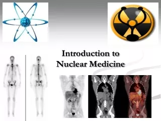
What is Nuclear Medicine * Inject radionuclide into the body. * Allow for distribution and uptake/metabolism of radionuclide (Radiopharmaceuticals). * A gamma camera detect gamma rays from the radionuclide after a certain time. * Image reconstruction.
There are two popular method in Nuclear Medicine * Positron Emission Tomography (PET). * Single photon emission computed tomography (SPECT)
Physics Explanations 1- A short lived radioactive isotope (18F), is injected into the living subject (usually into blood circulation ) 2- Host cells uptake radionuclide based on metabolic rate (there is a waiting period while the active molecule becomes concentrated in tissues of interest). 3- Radionuclide generates gamma ray. .
4- Gamma ray measured by Nuclear technique and record data. 5- Millions of these data points are collected and analyzed. 6- Nuclear technique can create an image of the required part (such as, the brain).
How to obtain a functional imaging?1- The patient is injected with radionuclide.
Image is strongly depended on uptake the amount of radionuclide in organ tissue.
History of Nuclear medicine • 1896 • Henri Becquerel discovered "rays" from uranium. • 1897 • Marie Curie named the rays "radioactivity“.
1925 • HerrmanBlumgart and Otto Yens used (radium isotope) to determine the velocity of blood.
1938 • JohnLivingood and Glenn Seaborg discovered iodine-131 and cobalt-60. • Emilio Segre and Glenn Seaborg discovered technetium-99m.
1951 • Benedict Cassen, Lawrence Curtis, and Raymond Libby used a scintillation detector to "scan" the distribution of radioiodine within the thyroid gland.
1958 • Hal Anger invented the scintillation camera, an imaging device that made it possible to conductdynamic studies.
1963 • Henry Wagner first used radio-labeled albumin aggregates for imaging lung in normal persons and patients.
1971 • The American Medical Association recognized that, nuclear medicine as a medical specialty. • 1983 • Henry Wagner carried out the first successful PET imaging of a neuroreceptor using himself as the experimental subject.
2008 • The first hybrid PET/MRI system for humans, created by Siemens, was installed.
Diagnostic Nuclear Medicine • Imaging (Planer/SPECT and PET Cameras) • Brain *Bone • Lungs *Thyroid • Kidneys *Liver/Spleen • Cardiovascular *Stomach • Tumors
Therapeutic Nuclear Medicine • Examples • Bone pain. • Thyroid cancer. • Lymphoma treatment.
Radiopharmaceutical • Radioactivity, is an element with unstable nucleus (excess energy)can emits radiation (α, β, γ). • When it is used in medical studies, named: RADIOPHARMACEUTICAL.
Radiation can be:- As a particle: α, β or as a photon: γ • Particles (α, β ) have mass, and can carry high energy in a short length. Therefore, radiopharmaceuticals with βdecay are used in therapy, also γ-ray used in therapy. • Photons (γ) has no mass, and can carry low energy in a long distance. Therefore, radiopharmaceuticals with γdecay are used in imaging.
Penetration radiation and non-penetration radiation (Gamma ray has high penetration)
Finally Radiopharmaceuticals Gives Radiation, which are used for the purpose of diagnosis or therapy, such as Iodine-131 (131I), Xenon-133 (133Xe) and Technetium 99mTc, etc.
Any materials consists of a large number of atoms or molecules ( each one gram of matter contains 6.022 X 10²³ atom). Such as, Iron, Wood, plastic, air and living tissues.
Reactor-produced radionuclide • According the following nuclear reaction, we can obtain the radionuclide (Isotope). A-1 ZX + n A-1ZX1 + γ X represent the nucleus, n is the neutron and X1 is radionuclide (produce γray).
Advantages of NM • Assesses body function and in monitoring flow rate of blood. • Early detection of pathology. (Stress fracture and tumors). • Whole body scanning is possible.
Disadvantages of NM • Radiation risks due to administered radionuclide. • Usually requiring an injection into the blood stream. • Disposal of radio activity waste, including that from patients, require special procedure. • Relatively high costs associated with radio tracer production and administration.