COX-2
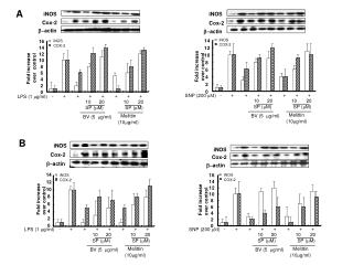
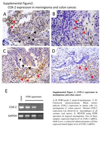
Supplemental Figure2. COX-2 expression in meningioma and colon cancer. B. A. D. C. E. Supplemental Figure 2. COX-2 expression in meningioma and colon cancer .

COX-2
E N D
Presentation Transcript
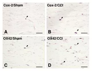
Supplemental Figure2. COX-2 expression in meningioma and colon cancer. B A D C E Supplemental Figure 2.COX-2 expression in meningioma and colon cancer. A, B; WHO grade 2, atypical meningioma. C, D; Colorectal adenocarcinoma. Black arrows indicate COX-2 expression in tumor cells (A; meningioma, C; colon cancer). Distinct COX-2 expression was observed in macrophages in all sections (red arrow). E: RT-PCR using FFPE specimen of atypical meningioma. Two of three samples expressed high-level of COX-2 mRNA, while Lovo, a colorectal adenocarcinoma cell line, expressed much higher level of COX-2 mRNA. FFPE specimen Lovo COX-2 GAPDH
Supplemental material and method RNA Isolation and RT-PCR Analysis Total RNA was isolated from formalin-fixed paraffin embedded (FFPE) specimens of three atypical meningioma following the manufacturer’s instructions using the RNeasyFFPE Kit(Quiagen, Hilden, Germany). Complementary DNA was synthesized by using reverse transcriptase (Superscript II, Invitrogen, Karlsruhe, Germany) and oligo (dT) primers, and used as template for PCR reactions. PCR was carried out using COX-2 primers and GAPDH primers. . Primers for COX-2 were: forward- 5’-ATGCTCGCCCGCGCCCTGCTGCT-3’, reverse- 5’-CCAGTATAAGTGCGATTGTACCCG-3’ and GAPDH were: forward- 5’-CGGGTACAATCGCACTTATACTGG-3’, reverse- 5’-GATGCAGGGATGATGTTC-3’ . A PCR mixture (total volume 25 μL) was prepared that included 2 μL of the sample containing each nucleotide, each primer at final concentration of 200 nM, and 9.5 uL of Taq-polymerase (Go Taq Green Master Mix, Promega, Madison, WI, USA). As a positive control of RT-PCR, the colon cancer cell line (Lovo) was employed. Each PCR reaction was run for 33 cycles with a denaturation step for 30 sec at 95C, and an extension for 60 seconds at 72 C, Annealing was for 30 seconds at 55 C.