AIMS
MATERIALS & METHODS. AIMS. RESULTS. 1 μ m. 0.2 µ m. 0.2 µ m. 4A. 3B. 3A. 4B. 0.2 µ m. 0.2 µ M. 0.5 µ m. 0.2 µ M. CONCLUSION

AIMS
E N D
Presentation Transcript
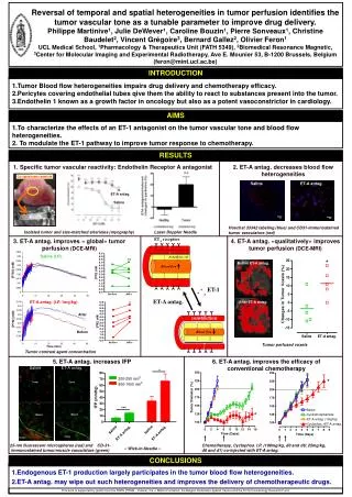
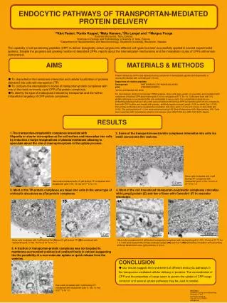
MATERIALS & METHODS AIMS RESULTS 1 μm 0.2 µm 0.2 µm 4A 3B 3A 4B 0.2 µm 0.2 µM 0.5 µm 0.2 µM CONCLUSION ●Our results suggest the involvement of different endocytic pathways in the transportan-mediated cellular delivery of proteins. The concentration of CPP and the properties of cargo seem to govern the uptake of CPP-cargo construct and severaluptake pathways may be used in parallel. ENDOCYTIC PATHWAYS OF TRANSPORTAN-MEDIATED PROTEIN DELIVERY1,2Kärt Padari, 2Kaida Koppel, 3Mats Hansen, 3Ülo Langel and 1,2Margus Pooga1Estonian Biocentre, Tartu, Estonia2Institute of Zoology and Hydrobiology, University of Tartu, Estonia3Department of Neurochemistry and Neurotoxicology, Stockholm University, Stockholm, Sweden The capability of cell penetrating peptides (CPP) to deliver biologically active cargoes into different cell types has been successfully applied in several experimental systems. Despite the progress and growing number of described CPPs, reports about the internalization mechanisms and the intracellular routes of CPPs still remain controversial. Protein delivery by CPPs was assessed usingcomplexes of biotinylated peptide and streptavidin or neutravidin labeled with colloidal gold (10 nm). Sequences of studied peptides Transportan: GWTLNSAGYLLGK*INLKALAALAKKIL pTat: G*RKKRRQRRRPQ *amino acid labeled with biotin For transmission electron microscopy (TEM) analysis, HeLa cells were grown on coverslips and incubated with complexes of biotinyl-CPP and protein-gold (10 nm) conjugateat 37˚C for 1 h. Cells were fixed with 3% glutaric aldehyde in cacodylate buffer and embedded in epoxy resin. For colocalization study by pre-embedding labeling method, HeLa cells were incubated with biotinyl-CPP and protein-gold (10 nm) complexes, fixed with PLP-fixative and treated with primary antibody against mouse Lamp2 (1:20) or rabbit Cav1 (1:50). The primary antibodies were visualized by incubation with nano-gold (1.4 nm) anti-mouse or anti-rabbit IgG (1:60). The gold particles of 1.4 nm were enhanced using HQ Silver Enhancement Kit (Nanoprobes, US). Cells were examined with transmission electron microscope(Jeol JEM-100S and JEM-1200 EXII, Japan). ●To characterize the membrane interaction and cellular localization of proteins delivered into cells with transportan (TP) ●To compare the internalization modes of transportan-protein complexes with one of the most commonly used CPP pTat-protein complexes. ●To identify the type of endocytosis induced by transportan and the further intracellular targeting of CPP-protein complexes. • The transportan-streptavidin complexes associate with • filopodia or shorter microspikes at the cell surface and internalize into cells • by induction of large invaginations of plasma membrane allowing to • speculate about the role of macropinocytosis in the uptake process. 2. Some of the transportan-neutravidin complexes internalize into cells via small caveosome-like vesicles. HeLa cells incubated with 3 μM biotinyl-TP complexed with neutravidin gold (1:100, 10 nm) at 37 °C for 1 h. HeLa cells incubated with 2.5 μM biotinyl-TP complexed with streptavidin gold (1:50, 10 nm) at 37 °C for 1 h. 3. Most of the TP-protein complexes are taken into cells in the same type of endocytic structures as pTat-protein complexes. 4. Most of the cell transduced transportan-neutravidin complexes colocalize with Lamp2 protein (E) and few of them with Caveolin1 (F) in vesicular structures. HeLa cells incubated with 3 μM biotinyl-Tat (3A) and3 μM biotinyl-TP (3B) complexed with neutravidin gold (1:100, 10 nm) at 37 °C for 1 h. HeLa cells incubated with 3 μM biotinyl-transportan complexed with neutravidin-gold (1:100, 10 nm) at 37 °C for 1 h. Cells were treated with primary antibody Lamp2 (4A) and Cav1 (4B) followed by incubation with secondary antibody labeled with nano-gold particles (1.4 nm). 5. A fraction of transportan-protein complexes was not targeted to membrane-surrounded vesicles but localized freely in cytosol suggesting the the possibility of a non-vesicular uptake or quick release from the vesicles. HeLa cells incubated with 3 μM biotinyl-TP complexed with neutravidin-gold (1:100, 10 nm) at 37 °C for 1 h. Kärt Padari Institute of Zoology and Hydrobiology, University of Tartu Vanemuise Street 46, Tartu 51014, ESTONIA kartp@ut.ee