Download
1 / 15
150 likes | 189 Vues
Explore the different tissues in fish thyroid and kidney, from normal to hyperplasia and depletion, understanding the physiological implications. Gain insights on melanomacrophage activity and its relation to haematopoietic changes in various fish species.

E N D
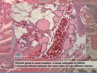
Thyroid gland in usual location– a loose collection of colloid containing follicles between the lower jaws of 5 gm Atlantic salmon.
The thyroid of this young salmon is forming a less diffuse organ.
The location of the normal (usual) thyroid can perhaps be better appreciated in this galaxid with goitre. (Yes, they do get goitre, particularly in freshwater from iodine deficient areas.) I have not recognized this in thyroid follicles within the kidney.
And another Ring-In: perivascular tissue in head kidney of an Angel Fish with skin lesions (ignore the necrosis). • What do you think this is? (Hint - mentally remove the haematopoietic tissue to long bones) Correct?You are left with cells round blood vessels anterior to the nephron-kidney that resemble those of the adrenal cortex and medulla - and are functionally the same. Termed inter-renal tissue (cortical equivalent) and chromaffin cells(medullary equivalent). Variations in the amount of inter-renal tissue may reflect area of section, physiological variations such as reproductive activity in some fish, or chronic stress. Note:frequently the distinction of these 2 cells types is not obvious in routine sections.
The effect of physiological state on the kidney interstitial tissue Changes in haematopoietic activityThis is often easier to assess in the tail or posterior kidney, where the nephrons help gage the amount of interstitial tissue present.
Posterior or tail kidney - normal young actively growing salmon with normal active haematopoiesis. … And that many of the haematic tissue cells how signs of maturation, such as a polymorphonuclear appearance. There are only occasional mitoses. Note the relative proportions of tubules and haematopoietic tissue
If we look at another area from the same kidney: Note occasional mitosis & mature cells e.g. granulocytes
Another example from this fish, illustrating that the distribution of interstitial tissue may not be uniform - need to consider the overview.
Haematopoietic Hyperplasia Increased haematopoietic activity in salmon smolt recently transferred to sea . Note high mitotic rate (arrows) and a dominance of immature cells including blast-like forms.
Hyperplasia is seen as an overall increase in interstitial cells seen both as high density - & slight separation of tubules. Mitoses ( ) Note both these animals are growing. Differences are only those of degree. “Smoltification” involves pre-adaptation to a marine environment. Animals entering the sea are readily able to regulate the high salt level and undergo a metabolic boost in thyroid & other hormones>increased growth>including increased HP activity.
Haematopoietic tissue depletion Haematopoietic tissue varies from sparse to normal, with some visible spaces (& a little tubule shrinkage artifact). In comparison, this salmon kidney shows closely packed tubules, with some spaces between. There is no evidence of cell loss due to cell death. Reduced interstitial tissue may reflect low levels of haematopoiesis (look for other indications of low growth & metabolic rate), but where there is an increase in tissue spaces this may also reflect mobilization of mature blood cells in response to stress or infections.
Another young salmon showing marked haematopoietic depletion, following (unidentified) stress. Only blood vessels separate many tubules.
And this one? No, not wildly depleted! This is normal. The fact that it has no haematopoietic tissue at all is the give-away. This is the tail kidney of a River Blackfish (Gadopsis marmoratus ) a species that has more complete separation of head kidney (haematopoietic tissue) and nephron containing tail kidney.
The effect of physiological state on the kidney interstitial tissue B. Changes in melanomacrophage activity.Increased melanomacrophage activity reflects phagocytic activity or tissue turnover. It may be accompanied by altered haematopoietic (HP) activity.For example, fish that are undergoing catabolism due to prolonged cessation of feeding will also show reduced HP activity.
Wild Brown trout: It is common to find more pigment in wild fish than similar aged farmed animals receiving a more concentrated & available diet & growing more rapidly. Judging whether this increase can be attributed to differences in diet or age, or reflect a pathological process of tissue turnover, is an interpretive skill requiring a good history to support histology findings.