Download
1 / 20
200 likes | 222 Vues
AFNI is a software developed for FMRI data analysis, offering a wide range of tools and features. This article introduces the concepts and principles behind AFNI and provides an overview of its functionalities and capabilities.

E N D
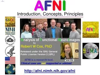
AFNI &FMRIIntroduction, Concepts, Principles http://afni.nimh.nih.gov/afni
AFNI = Analysis of Functional NeuroImages • Developed to provide an environment for FMRI data analyses • And a platform for development of new software • AFNI refers to both the program of that name and the entire package of external programs and plugins (more than 200) • Important principles in the development of AFNI: • Allow user to stay close to the data and view it in many different ways • Give users the power to assemble pieces in different ways to make customized analyses • “With great power comes great responsibility” — to understand the analyses and the tools • “Provide mechanism, not policy” • Allow other programmers to add features that can interact with the rest of the package
Principles (and Caveats) We* Live By • Fix significant bugs as soon as possible • But, we define “significant” • Nothing is secret or hidden (AFNI is open source) • But, possibly not very well documented or advertised • Release early and often • All users are beta-testers for life • Help the user (message board; consulting with NIH users) • Until our patience expires • Try to anticipate users’ future needs • What we think you will need may not be what you actually end up needing *
Before We Really Start • AFNI has many programs and they have many options • Assembling the programs to do something useful and good seems confusing (OK, isconfusing) when you start • To help overcome this problem, we have “super-scripts” that carry out important tasks • Each script runs multiple AFNI programs • We recommend using these as the basis for FMRI work • When you need help, it will make things simpler for us and for you if you are using these scripts • afni_proc.py= Single subject FMRI pre-processing and time series analysis for functional activation • uber_subject.py = GUI for afni_proc.py • align_epi_anat.py = Image alignment (registration), including anatomical-EPI, anatomical-anatomical, EPI-EPI, and alignment to atlas space (Talairach/MNI)
D: 4-5 s rise E: 5 s plateau C: ≈ 2 s delay B:5 s neural activity F: 4-6 s fall A What is Functional MRI? • 1991: Discovery that MRI-measurable signal increases a few % locally in the brain subsequent to increases in neuronal activity (Kwong, et al.) Signal increase caused by change in H2O surroundings: more oxygenated hemoglobin is present Cartoon of MRI signal in a single “activated” brain voxel Contrast through time with no noise! G: Return to baseline (or undershoot) A: Pre-activation baseline time
How FMRI Experiments Are Done • Alternate subject’s neural state between 2 (or more) conditions using sensory stimuli, tasks to perform, ... • Can only measure relative signals, so must look for changes in the signal between the conditions • Acquire MR images repeatedly during this process • Search for voxels whose NMR signal time series (up-and-down) matches the stimulus time series pattern (on-and-off) • FMRI data analysis is basically pattern matching in time • Signal changes due to neural activity are small • Need 500 or so images in time series (in each slice) takes 30 min or so to get reliable activation maps • Usually break image acquisition into shorter “runs” to give the subject and scanner some break time • Other small effects can corrupt the results post-process the data to reduce these effects & be vigilant • Lengthy computations for image recon and temporal pattern matching data analysis usually done offline
Sample Data Time Series • 64×64 matrix (TR=2.5 s; 130 time points per imaging run) • Somatosensory task: 27 s “on”, 27 s “rest” • Note that this is really good data pattern of expected BOLD signal pattern fitted to data data One echo-planar image One anatomical image, with voxels that match the pattern given a color overlay
Fundamental AFNI Concepts • Basic unit of data in AFNI is the dataset • A collection of 1 or more 3D arrays of numbers • Each entry in the array is in a particular spatial location in a 3D grid (a voxel= 3D pixel) • Image datasets: each array holds a collection of slices from the scanner • Each number is the signal intensity for that particular voxel • Derived datasets: each number is computed from other dataset(s) • e.g., each voxel value is a t-statistic reporting “activation” significance from an FMRI time series dataset, for that voxel • Each 3D array in a dataset is called a sub-brick • There is one number in each voxel in each sub-brick Jargon! Jargon! } 3x3x3 Dataset With 4 Sub-bricks
What's in a Dataset: Header • Besides the voxel numerical values, a dataset also contains auxiliary information, including (some of which is optional): • xyz dimensions of each voxel (in mm) • Orientation of dataset axes; for example, x-axis=R-L, y-axis=A-P, z-axis=I-S = axial slices (we call this orientation “RAI”) • Location of dataset in scanner coordinates • Needed to overlay one dataset onto another • Very important to get right in FMRI, since we deal with many datasets • Time between sub-bricks, for 3D+time datasets • Such datasets are the basic unit of FMRI data (one per imaging run) • Statistical parameters associated with each sub-brick • e.g., a t-statistic sub-brick has degrees-of-freedom parameter stored • e.g., an F-statistic sub-brick has 2 DOF parameters stored Jargon!
AFNI Dataset Files - 1 • AFNI formatted datasets are stored in 2 files • The .HEAD file holds all the auxiliary information • The .BRIK file holds all the numbers in all the sub-bricks • Datasets can be in one of 3 2 coordinate systems (“views”) • Original data or +orig view: from the scanner • AC-PC aligned or +acpc view: • Dataset rotated/shifted so that the anterior commissure and posterior commissure are horizontal (y-axis), the AC is at (x,y,z)=(0,0,0), and the hemispheric fissure is vertical (z-axis) • Talairach or +tlrc view: • Dataset has also been rescaled to conform to the Talairach-Tournoux atlas dimensions (R-L=136 mm; A-P=172 mm; I-S=116 mm) • AKA Talairach or Stererotaxic coordinates • Not quite the same as MNI coordinates, but very close • Actually, all datasets scaled+aligned to an atlas are labeled +tlrc • Header can contain name of actual atlas “space”
AFNI Dataset Files - 2 • AFNI dataset filenames consist of 3 parts • The user-selected prefix (almost anything) • The view (one of +orig, +acpc, or +tlrc) • The suffix (one of .HEAD or .BRIK) • Example: BillGates+tlrc.HEAD and BillGates+tlrc.BRIK • When creating a dataset with an AFNI program, you supply the prefix; the program supplies the rest • AFNI programs can read datasets stored in several formats • ANALYZE (.hdr/.img file pairs); i.e., from SPM, FSL • MINC-1 (.mnc); i.e., from mnitools • CTF (.mri, .svl) MEG analysis volumes • ASCII text (.1D) — numbers arranged into columns • Have conversion programs to write out MINC-1, ANALYZE, ASCII, and NIfTI-1.1 files from AFNI datasets, if desired Jargon!
NIfTI Dataset Files • NIfTI-1.1 (.niior.nii.gz) is a standard format that AFNI, SPM, FSL, BrainVoyager, et al., have agreed upon • Adaptation and extension of the old ANALYZE 7.5 format • Goal: easier interoperability of tools from various packages • All data is stored in 1 file (cf. http://nifti.nimh.nih.gov/) • 348 byte header (extensions allowed; AFNI uses this feature) • Followed by the image binary numerical values • Allows 1D–5D datasets of diverse numerical types • .nii.gz suffix means file is compressed (with gzip) • AFNI now reads and writes NIfTI-1.1 formatted datasets • To write: when you give the prefix for the output filename, end it in “.nii” or “.nii.gz”, and all AFNI programs will automatically write NIfTI-1.1 format instead of .HEAD/.BRIK • To read: just give the full filename ending in “.nii” or “.nii.gz”
Getting and Installing AFNI • AFNI runs on Unix systems: Linux, Sun, Mac OS X • Can run under Windows with Cygwin Unix emulator • This option is really just for trying it out — not for production use! • You can download precompiled binaries from our Website • http://afni.nimh.nih.gov/afni • Also: documentation, message board, humor, data, class materials, … • You can download source code and compile it • Also from GitHub: https://github.com/afni/AFNI • AFNI is updated fairly frequently, so it is important to update occasionally -- @update.afni.binaries • We can’t help you with outdated versions! • Please check for updates every 6 months (or less)
AFNI at the NIH Scanners • AFNI can take 2D images in “realtime” from an external program and assemble them into 3D+time datasets slice-by-slice • FMRI Facility scanners at the NIH (GE and Siemens) are set up to start AFNI on a remote Linux computer automatically when EPI acquisition starts, and then the Dimon program is used to send images into AFNI as they are reconstructed: • For immediate display (images and graphs of time series) • Plus: graphs of estimated subject head movement • Goal is to let you see image data as they are acquired, so that if there are any big problems, you can fix them right away • Sample problem: someone typed in the imaging field-of-view (FOV) size wrong (240 cm instead of 24 cm), and so got garbage data, but only realized this too late (after scanning 8 subjects this way) — D’oh!
Other Parts of AFNI • Batch mode programs and scripts • Are run by typing commands directly to computer, or by putting commands into a text file (script) and later executing them • Good points about batch mode • Can process new datasets exactly the same as old ones • Can link together a sequence of programs to make a customized analysis (a personalized pipeline) • Some analyses take a long time (are not interactive) • Bad points about batch mode • Learning curve is “all at once” rather than gradual • If you are, like, under age 35, you may not know how to, like, type commands into a computer to make it do things • But we don’t make you use punched cards or paper tape (yet)
AFNI Batch Programs • Many many important capabilities in AFNI are onlyavailable in batch programs • A few examples (of more than 100, from trivial to complex) • 3dDeconvolve+3dREMLfit= multiple linear regression on 3D+time datasets; fits each voxel’s time series to activation model, tests these fits for significance (3dNLfim= nonlinear fitting) • 3dvolreg = 3D+time dataset registration, to correct for small subject head movements, and for inter-day head positioning • 3dANOVA + 3dLME = 1-, 2-, 3-, and 4- way ANOVA/LME layouts: combining & contrasting datasets in Talairach space • 3dcalc = general purpose voxel-wise calculator (very useful) • 3dsvm = SVM multi-voxel pattern analysis program • 3dresample = re-orient and/or re-size dataset voxel grid • 3dSkullStrip = remove “skull” from anatomical dataset • 3dDWItoDT = compute diffusion tensor from DWI (nonlinearly)
SUMA, et alii • SUMA is the AFNI surface mapper • For displaying surface models of cortex • Surfaces from FreeSurfer (MGH) or Caret (Wash U) or BrainVoyager (Brain Innovation) • Can display functional activations mapped from 3D volumes to the cortical surface • Can draw ROIs directly on the cortical surface • vs. AFNI: ROIs are drawn into the 3D volume • SUMA is a separate program from AFNI, but can “talk” with AFNI (like a plugout) so that volume & surface viewing are linked • Click in AFNI or SUMA to change focus point, and the other program jumps to that location at the same time • Functional (color) overlay in AFNI can be sent to SUMA for simultaneous display • And much more — stayed tuned for the SUMA talks to come!
SUMA Teaser Movie Color from AFNI, Images from SUMA Images captured with the ‘R’ recorder function, then saved as animation with Save:aGif control
Other Educational Presentations • How to get images into AFNI or NIfTI format (program to3d) • Detailed hands-on with using AFNI for data viewing (fun) • Signal modeling & analysis: theory & hands-on (3dDeconvolve et al.) • Image registration (3dvolreg et al.) • Volume rendering hands-on (fun level=high) • ROI drawing hands-on (fun level=extreme) • Transformation to Talairach hands-on (fun level=low) • Group analysis: theory and hands-on (3dANOVAxand beyond) • Experiment design • FMRI analysis from start to end (the “soup to nuts” hands-on) • SUMA hands-on (fun level=pretty good) • Surface-based analysis • Connectivity (resting state, white matter tracts) • AFNI “Jazzercise”(practice sessions & directed exercises)