Download
1 / 49
510 likes | 766 Vues
Cerebral Palsy. CEREBRAL PALSY Diagnostic term used to describe a group of motor syndromes resulting from disorders of early brain development . Symptom complex , (not a disease ) that has multiple etiologies. Brain damage Occurs during developmental period Motor dysfunction

E N D
CEREBRAL PALSY Diagnostictermusedtodescribe a group of motor syndromes resultingfromdisorders of earlybraindevelopment. Symptomcomplex, (not a disease) that has multipleetiologies.
Braindamage Occursduringdevelopmentalperiod Motor dysfunction Not Curable Non-progressive (static) Anyregressionordeterioration of motor orintellectualskillsshouldprompt a searchfor a degenerativedisease Therapy can helpimprovefunction
CP is causedby a broadgroup of Developmental GeneticProduce a common Metabolicgroup of neurologicphenotypes Ischemic Infectious Otheracquiredetiologies
CP is associatedwith Epilepsy Abnormalitiesspeech, vision, intellect Selectivevulnerability of thebrain’s motor system Manychildrenandadultsfunction at a higheducationallevel
There are 2 major types of CP, depending on location of lesions: • Pyramidal (Spastic) • Extrapyramidal • There is overlap of both symptoms and anatomic lesions.
Types of brain damage • Bleeding • Brain malformation • Trauma to brain • Lack of oxygen • Infection • Toxins • Unknown
Etiology Antenatalfactorscausingabnormalbraindevelopment Congenitalanomalies Intrapartumasphyxia Intrauterineexposuretomaternalinfection Multiplebirths Lowbirthweightinfants Intracerebralhemorrhage Periventricularleukomalacia
Hypoxic Ischemic Encephalopathy (HIE) • A clinical entity first described in 1976 • Used interchangeably with Neonatal encephalopathy. • Asphyxia refers to the first minutes after birth (low Apgars and acidosis) • HIE signs and symptoms persist over hours and days that follow.
Hypoxic Ischemic Encephalopathy (HIE) 3 major lesions arise from HIE • PeriventricularLeukomalacia (PVL) Typically seen in the premature infant a. Hemorrhagic PVL b. Ischemic PVL • Parasaggital Cerebral Injury Typically seen in the term infant • Selective (Focal) Neuronal Necrosis Seen in both term and premature infants
Periventricular Leukomalacia (PVL) Hemorrhagic PVL Periventricular venous congestion (swelling)mayoccur, and cause ischemia (lack of blood supply) and periventricular hemorrhagic infarction.
Periventricular Leukomalacia (PVL) Ischemic PVL • An ischemic infarction or failure of perfusion usually to the watershed area surrounding the ventricular horns- “HIE white matter necrosis”. • Peak incidence occurs around 32 weeks • Larger infarcts may leave a cyst • Secondary hemorrhage can occur into theses cysts- “periventricular hemorrhage”.
Periventricular Leukomalacia (PVL) Ischemic PVL • PVL can extend into the internal capsule and result in hemiplegia superimposed on diplegia. • Prenatal maternal ultrasound has detected lesions in the fetus at 28-32 weeks gestation, thus confirming that PVL can occur prenatally.
Parasaggital Cerebral Injury • Injury is related to vascular factors, especially in the parasaggital border zones that are more vulnerable to a drop in perfusion pressure and immature autoregulation. • The ischemic lesion results in cortical and subcortical white matter injury. • It is usually bilateral and symmetric. • The posterior aspect of the cerebral hemisphere especially the parietal occipital regions is more affected than the anterior.
Selective (Focal) Neuronal Necrosis (SNN) • Occurs in the glutamate sensitive areas in the basal ganglia, thalamus, brainstem and cortex. • The location of the focal necrosis, which show up as cystic lesions on MRI, depend on the stage of development of the infant’s brain at the time of the HIE. • For example, HIE at term often produces SNN in the basal ganglia since it is glutamate sensitive and very hypermetabolic at term.
Pyramidal Velocity dependent increased resistance to passive muscle stretch The spasticity can be worse when the person is anxious or ill. The spasticity does not go away when the person is asleep. Extrapyramidal Ataxia Hypotonia Dystonia Rigidity The tone may increase with volitional movement, or when the person is anxious During sleep the person is actually hypotonic Types of Cerebral Palsy
Types of Cerebral Palsy Pyramidal (Spastic) • Quadriplegia- all 4 extremities • Hemiplegia- one side of the body • Diplegia- legs worse than arms • Paraplegia- legs only • Monoplegia- one extremity
Dyskinetic Athetosis Chorea- quick, jerky movements Choreoathetosis- mixed Hypotonia- floppy, low muscle tone, little movement Ataxic CP Results from damage to the cerebellum Ataxia- tremor & drunken- like gait ExtrapyramidalDivided into Dyskinetic and Ataxic types
Pyramidal Lesion is usually in the motor cortex, internal capsule and/or cortical spinal tracts. Extrapyramidal Lesion is usually in the basal ganglia, Thalamus, Subthalamic nucleus and/or cerebellum. Anatomy
SPASTIC DIPLEGIA Periventrıcularleukomalacia (PVL) Prematurity Ischemia Infection Endocrine /Metabolic
SPASTIC QUADRIPLEGIA PVL Multicysticencephalamalacia Malformation infection endocrine/ metabolic genetic/ developmental
HEMIPLEGIA Strokeinuteroorneonatal Thrombophylicdisorders Infection Genetic/ developmental Periventricularhemorrhage - infection
EXTRAPYRAMIDAL (athetoid-dyskinetic) Pathology,putamen,globuspallidus,thalamus, basalganglia Asphyxia Kernicterus Mitochondrial Genetic/metabolic
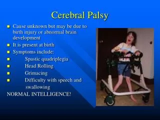
SYMPTOMS All types of CP are characterized by Abnormal muscle tone Reflexes Motor development Coordination
ClassicalSymptoms Spasticities Spasms Involuntarymovements Unsteadygait Problemswithbalance Scissorwalking Toewalking
Babiesbornwith severe CP oftenhave an irregularposture floopyorstiff spinalcurvature smalljawbone
SPASTIC HEMIPLEGA Decreasedspontaneousmovements on theaffectedside Thearm is oftenmoreinvolvedthantheleg Difficulty in handmanipulation is obviousby 1 yr of age Walking is delayeduntil 18-24 months Circumductivegait is apparent
Examination of theextreminitesmayshowgrowtharrest Spasticity is apparent in theaffectedextremities An affectedchildoftenwalks on tiptoe Ankleclonusand a Babinskisignmay be present DTR areincreased
1/3 of patientshave a seizuredisorder 25% havecognitiveabnormalities
CT or MRI →An atrophiccerebralhemispherewith a dilatedventriculecontrlateraltotheside of theaffectedextremities CT →Usefulfordetectingcalcificationsassociatedwithcongenitalinfections Familyhistoriessuggestive of thrombosisandinheritedclottingdisordersmay be present
SPASTIC DIPLEGIA Themostcommon form of thespasticforms Bilateralspasticity of thelegs Firstindication is oftennotedwhen an infantbeginstocrawl = commandocrawl Ifthespasticity is severe application of diaper is difficult
Ankleclonus, Babinskisign ( bilateral) Scissoringposture of thelowerextremities Walking is delayed Childwalks on tiptoe Impairedgrowth of lowerextremities Hipproblems,dislocations, strabismus Normal intellectualdevelopment
SPASTIC QUADRIPLEGIA → ( TETRAPLEGIA) Most severe form of CP Motor impairment of all extremities High association with mental retardation and seizures Swallowing difficulties are common → aspiration pneumonia Increased tone and spasticity Brisk reflexes, plantar extensor responses Speech and visual abnormalities
ATHETOID CP= EXTRAPYRAMIDAL CP Lesscommon Affectedinfantsarecharacteristicallyhypotonicwithpoorheadcontrol Developedincreasedvariabletonewithrigidityanddystoniaoverseveralyears Feedingmay be difficult
Speech is typicallyaffected Oropharyngenalmusclesareinvolved Seizuresareuncommon Can also be causedbykernicterus
DIAGNOSIS History Physical examinaton Neurological examination MRI → determine the location and extent of structural lesions,associated congenital anomalies Hearıng and visual function test Genetic evaluation
TREATMENT Multidisciplinary approach in the treatment Physians from various specialities Occupational and physical therapist Speech pathologist Social workers Educators Developmental psychologist
Parents should be taught now to work with their children in daily activities Feeding Carrying Dressing Bathing Playing Need to be instructed in the supervision of a series of exercises to prevent the development of the contractures
Spastic diplegia → treated initially with the assistance of adaptive equipment such as walkers some surgical procedures that reduce muscle spasm.
Quadriplegıa Motorized wheelchairs Special feeding devices Modified typewriters Customized seating arrangements
Hemiplegia Improved hand or arm functioning on the affected side
Orthopedic Problems • Scoliosis • Hip Dislocations • Contractures • Osteoporosis
Medical Management Oromotor Dysfunction • Especially common in persons with Extrapyramidal CP and Spastic quadriplegia • Language delay/Speech delays • Drooling • Dysphagia • Aspiration
Medical Management Gastrointestinal Dysmotility • Delayed gastric emptying • Gastroesophageal reflux • Pain • Chronic aspiration • Constipation .
Medical Management Gastrointestinal Dysmotility • Delayed gastric emptying • Gastroesophageal reflux • Pain • Chronic aspiration • Constipation These disorders are interrelated and compound one another.
Medical Management Spasticity Management Management of spasticity does not fix the underlying pathology of CP, but it may decreased the sequelae of increased tone. • Over time, the spasticity leads to: • musculoskeletal deformity • scoliosis • hip dislocation • contractures • Pain • Hygiene problems
Treatment of Spasticity Medications • Valium • Dantrium • Baclofen • Clonidine • Clonazepam • BOTOX
Mental Retardation Communication Disorders Neurobehavioral Seizures Vision Disorders Hearing loss Somatosensation (skin sensation, body awareness) Temperature instability Nutrition Drooling Dentition problems Neurogenic bladder Neurogenic bowel Gastroesophageal reflux Dysphagia Autonomic dysfunction Associated Problems