Download
1 / 15
160 likes | 380 Vues
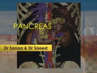
Anatomy of Pancreas. Yuniarti Anatomy department Faculty of Medicine UNISBA. Location of pancreas : Pancreas is an elongated, accessory digestive gland that lies retroperitoneally Transversely across the posterior abdominal wall posterior to the stomach between

E N D
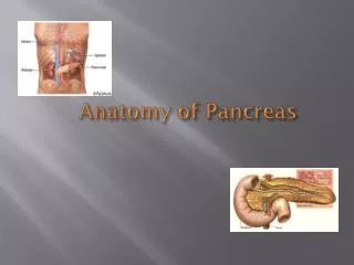
Anatomy of Pancreas Yuniarti Anatomy department Faculty of Medicine UNISBA
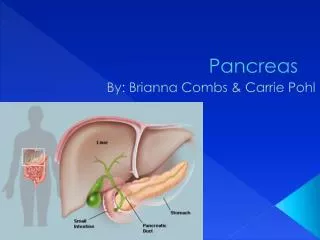
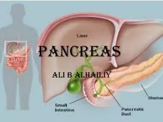
Location of pancreas : Pancreas is an elongated, accessory digestive gland that lies retroperitoneally Transversely across the posterior abdominal wall posterior to the stomach between duodenum on the right and the spleen on the left.
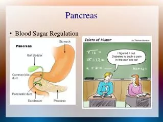
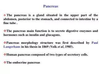
The pancreas produce : • An exocrine secretion ( pancreatic juice from the acinar cells) • that enter the duodenum through the main and accessory • pancreatic ducts. • Endocrine secretion (glucagon & insulin) from • the pancreatic islets (of langerhans) that enter blood.
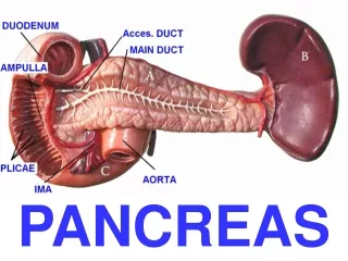
Parts of pancreas : • Head • Neck • Body • Tail
HEAD • The expanded part of the gland that is embraced by the C shaped curve of the duodenum to the right of the superior mesenteric vessels. • Firmly attaches to the medial aspect of the descending and horizontal parts of the duodenum. • The head of the pancreas rests posteriorly on the IVC • On its way to opening into the descending part of the duodenum, the bile duct lies in a groove on the posterosuperior surface of the head or is embedded in its substance.
NECK • Short and overlies the superior mesenteric vessels, which form a groove in its posterior aspect. • The anterior surface of neck, covered with peritoneum, is adjacent to the pylorus of the stomach.
BODY • Continues from the neck and lies to the left of the superior mesenteric vessels, • passing over the aorta and L2 vertebra • The posterior surface of the body is devoid of peritoneum and is in contact • with the aorta, SMA, left suprarenal gland and left kidney and renal vessels
TAIL • Lies anterior to the left kidney, where it is closely related to the left kidney,where it is closely related to the splenic hilum and the left colic flexure. • The tip of the tail is usually blunted and turned superiorly.
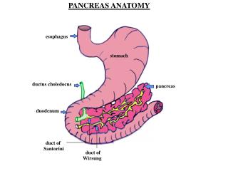
The main pancreatic duct • Begins in the tail of the pancreas and runs through the parenchyma of the gland • to the pancreatic head, here it turns inferiorly and is closely related to the bile duct. • Most of the time, the main pancreatic duct and the bile duct unite to form the short, • dilated hepatopancreatic ampulla (of vater), which opens into the descending part • of the duodenum at the summit of the major duodenal papilla
Accessory pancreatic duct • Opens into the duodenum at the summit of the minor duodenal papilla • Usually (60%) communicates with the main pancreatic duct
Smooth muscle sphincter that control the flow of bile & pancreatic juice • into duodenum : • - Sphincter of the pancreatic duct • - Sphincter of the bile duct • - Sphincter of hepatopancreatic (sphincter of Oddi)
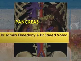
-The pancreatic arteriesderive mainly from the branches of the splenic artery -The anterior and posterior superior pancreaticoduodenal arteries, branches of the gastroduodenal artery -The anterior and posterior inferior pancreaticoduodenal arteries, branches of the SMA
The pancreatic lymphatic vessels follow the blood vessel. Most of them end in the pancreaticosplenic nodesthat lie along the splenic artery, but some vessels end in the pyloric lymph nodes. Efferent vessels from these nodes drain to the superior mesenteric lymph nodesor to the celiac lymph nodesvia the hepatic lymph nodes.
The nerves of the pancreas are derived from the vagus and abdominopelvic splanchnic nerves.