Respiratory and Circulatory Functions
610 likes | 1.24k Vues
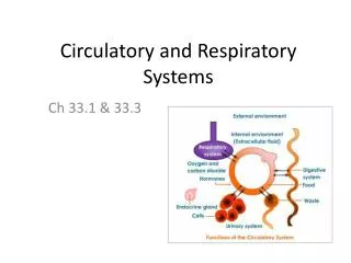
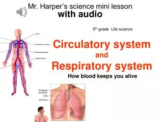
Respiratory and Circulatory Functions. Sections 30.1 – 30.2. Circulatory and Respiratory Systems. They work together to help maintain homeostasis in the body. What do they help to regulate? Body temp Heart rate Breathing rate O 2 and CO 2 levels in cells.

Respiratory and Circulatory Functions
E N D
Presentation Transcript
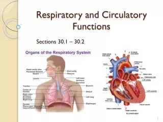
Respiratory and Circulatory Functions Sections 30.1 – 30.2
Circulatory and Respiratory Systems They work together to help maintain homeostasis in the body. What do they help to regulate? Body temp Heart rate Breathing rate O2 and CO2 levels in cells
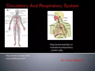
Function of Respiratory and Circulatory Systems Main job of Respiratory System Transport gases to and from the circulatory system What gases? Get oxygen (O2) into the body and remove waste gases (CO2) out of the body Main job of Circulatory System Moves blood to all parts of the body. Get oxygen (O2) into the body and remove waste gases (CO2) out of the body These systems work together!
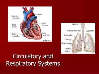
Respiratory and Circulatory Systems Components of Respiratory Nose Mouth Sinus Trachea Lungs Diaphragm • Components of Circulatory • Heart • Blood • Blood Vessels • Arteries • Veins • Capillaries
Pathway of Air into the Body Nose and Mouth-external opening to allow entry Air is filtered, cleaned, warmed, moistened Enters a series of tubes Protected by cartilage to keep tubes firm/open Mucus--traps foreign particles Cilia-- “sweep” foreign material away from lungs to be swallowed (or spit/coughed)
Structure of Alveoli • Small air sacs covered in mucus. • Wrapped in tiny blood vessels called capillaries
Respiratory System = system of tubes Trachea--main passageway to lungs Bronchi--two branches off the trachea that lead to lungs Bronchioles--smaller branches of the bronchi that lead to alveoli Alveoli--small air sacs at end of bronchioles 300 to 600 million in a pair of lungs Great deal of surface area Actual site of gas exchange
Breathing Mechanisms Involves muscles of the rib cage and the diaphragm Diaphragm: dome shaped muscle at the base of the rib cage Inhale: muscles contract, expand rib cage, diaphragm moves down Exhale: muscles relax, contract rib cage, diaphragm moves up
Breathing Mechanisms Air inhaled Air exhaled Rib cage lowers Rib cage rises Diaphragm Diaphragm Exhalation Inhalation
Gas Exchange 3 principles for gas exchange: O2 and CO2 are carried by the blood Gas moves by diffusion (moves from area of high to low concentration) Lining of the alveoli must be moist to help gases diffuse
Diffusion of gases Gases always move from areas of high concentration to areas of low concentration O2 concentration is higher in alveoli than blood: oxygen diffuses into blood At body cells O2 concentration is higher in blood: oxygen diffuses out of blood and into cells
Gas Exchange Oxygen alveoli capillaries red blood cells Carbon Dioxide red blood cells capillaries alveoli
Oxygen/Carbon Dioxide Transport O2 diffuses from alveoli to capillaries. O2 rich blood travels to heart and pumped to the body In tissues, O2 levels are lower, so RBCs release O2 to cells. In tissues, CO2 levels are higher, so CO2 diffuses from cells to blood. CO2 travels in blood to heart in form of Bicarbonate ions (HCO3-) Heart pumps blood to lungs where HCO3- breaks down to CO2 and water and then expelled out of body.
Circulatory System in detail Sections 30.3 – 30.5
Circulatory System Functions Carry O2 to cells and CO2 away from cells Deliver nutrients through body (after absorption in small intestine) Carry wastes away from cells (H2O, salt, urea) Help in fighting infections Temperature regulation
Parts of Circulatory System Heart: muscular pump that keeps blood moving throughout the system Blood: contains fluids, red blood cells, white blood cells, and platelets Blood vessels: carry blood to all of the body cells
Types of Blood Vessels Arteries: carry blood away from heart Veins: carry blood back to heart Capillaries: tiny blood vessels that deliver O2/remove CO2 from body cells
The Heart The heart is a muscular pump containing 4 chambers Made of special cardiac muscle that does not tire, unlike other muscles 2 smaller chambers at the top called atria (singular atrium) 2 larger chamber at the bottom called ventricles There are valves separating all of the chamber that prevent backflow
The Heartbeat Consists of 2 contractions Sinoatrial (SA) node sends electrical signal, which causes both of the atria to contract Electric signal spreads to the atrioventricular (AV) node AV node stimulates both ventricles to contract
Blood flow through the heart Into vena cava Into right atrium Down to right ventricle Out pulmonary artery to lungs Gets O2 and dumps CO2 Back to heart through pulmonary vein Into left atrium Down to left ventricle Out aorta to body
Two main circuits Pulmonary Blood goes from heart to lungs to pick up oxygen and release carbon dioxide Systemic Blood pumped out of heart to the rest of the body Sound of heart (lub/dub) made by valves closing
Blood flow through the body Blood vessels Arteries--take oxygenated blood away from the heart Thick/muscular and flexible walls Do not contain valves High Pressure
Blood flow through the body Capillaries Very small vessels Gases diffuse across very thin wall of small vessels Nutrients and oxygen leave the blood and go into the body tissue
Blood flow through the body Blood vessels Veins--take deoxygenated blood back to the heart Thin walls Larger diameters than arteries Have one-way valves Low Pressure
Circulation and Blood Pressure Blood Pressure: the forces with which blood pushes against the wall of an artery Healthy BP is 120/70 Systolic is top number, when the left ventricle contracts Diastolic is bottom number, when the left ventricle relaxes What could cause high BP? Blocked arteries Less elastic arteries Stress, high activity
Function: Delivery of nutrients and removal of wastes Blood delivers O2 to cells Delivers nutrients from digestion to cells (sugars, proteins) Blood takes CO2 back to lungs Delivers salts, water, and nitrogenous wastes (urea) to the kidneys for excretion Urea Main nitrogenous waste of body Produced in liver (from ammonia--NH3) Removed by kidneys
Function: Homeostasis (Maintain Temperature) In cold environments, blood vessels constrict in extremities to keep the torso and brain warm. In hot environments, blood vessels dilate and release excess heat.
Function: Homeostasis (Maintain Oxygen Levels) When cells need more oxygen, sensors in the walls of major arteries send signals to brain stem (medulla) Medulla coordinates with respiratory and circulatory systems to increase oxygen flow to cells and maintain homeostasis
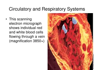
Components of Blood Blood contents Plasma 90% Water Nutrients, Proteins Wastes, Dissolved CO2 Cells Red blood cells (erythrocytes) Made in bone marrow 25 trillion of them (1000 RBC:1 WBC) Carry O2 and CO2 attached to protein called hemoglobin White blood cells (leukocytes) fight infection About 0.1% of all blood cells Platelets--involved in the formation of clots (along with fibrinogen (protein in blood) and blood cells)
ABO Basics • Blood group antigens are actually sugars attached to the red blood cell. • Antigens are “built” onto the red blood cell. • Individuals inherit a gene which codes for specific sugar(s) to be added to the red cell. • The type of sugar added determines the blood group. • If you need blood, you must get the same blood type or something compatible. • Incompatible blood will cause an immune response that can block vessels and could cause death.
ABO Blood Types Four blood types: A, B, AB, & O O has no protein markers A has A protein markers B has B protein markers AB has A and B protein markers
ABO Basics • The immune system produces an antibody (protein) in the plasma (produced by white blood cells) for the antigen not present. • Antibodies recognize foreigners and destroy them. Antibody B destroys antigen B. • For example, blood type A has antigen A attached to the red blood cell and antibody B in their plasma. Therefore, if blood type B is injected into their systems, anti-B antibodies in their plasma will recognize it as an alien and destroy it.
Rh Factor • Another important antigen (antigen D) found on the surface of red blood cells (RBCs) is the Rh factor. • Blood containing this antigen is said to be Rh positive (Rh+); blood lacking this antigen is said to be Rh negative (Rh-). • Positive can receive + or – blood • Negative can only receive – blood
RhDisease of the Neborn – How it Occurs • A) child is Rh+ and mother is Rh - • B) during pregnancy fetal Rh+rbc’s escape into maternal circulation • C) This causes the mother's immune system to make antibodies against the baby's red blood cells Rh (D) in future pregnancies. • D) Second pregnancy with Rh (D) pos child results in destruction of fetal D pos rbcs • This antibody response is called Rh sensitization and, depending on when it happens, can destroy the red blood cells of the baby before or after it is born. • If sensitization happens, a fetus or newborn can develop mild to severe problems (called Rh disease). In rare cases, if Rh disease is not treated, the fetus or newborn may die. • A woman with Rh-negative blood can get a shot of Rh immunoglobulin (such as RhoGAM) that almost always stops sensitization from occurring. Problems from Rh sensitization have become very rare since Rh immunoglobulin was developed.
Hemolysis If an individual is transfused with an incompatible blood group destruction of the red blood cells will occur. This may result in the death of the recipient.
Clumping • If a film remains uniform in appearance, there is no agglutination (clumping). • If the sample appears granular, agglutination has occurred.
When does the blood react? • Blood reacting to anti-A is group A. • Blood reacting to anti-B is group B. • Blood reacting to both anti-A and anti-B is group AB. • Blood not reacting to either anti-A or anti-B is group O. • Blood reacting to anti-Rh (D) is Rh+; Blood not reacting to anti-Rh (D) is Rh-
Blood Transfusions Blood must match for both ABO and Rh factor What type(s) of blood can AB- receive? What type(s) of blood can B+ receive? What type(s) of blood can O- receive?
How common is your blood type? 46.1% 38.8% 11.1% 3.9%