
Physiology of Vision: Depth Perception & Lens Accommodation | Dr. Sumera Gul
E N D
Presentation Transcript
Physiology of Vision Depth Perception and IOF Dr. Sumera Gul
Learning Objectives • At the end of the lecture the students should be able to: • Explain the mechanism of accommodation and its effects on the image formation • Discuss the determination of distance by depth perception • Explain the formation and flow of ocular fluid • Describe glaucoma and cataract
Refraction • This bending of light rays at an angulated interface is known as refraction. • Note particularly that the degree of refraction increases as a function of • (1) the ratio of the two refractive indices of the two transparent media and • (2) the degree of angulation between the interface and the entering • wave front.
Convex Lens Focuses Light Rays. focal point
Reduced Eye In the reduced eye, a single refractive surface is considered to exist, Its central point 17 millimeters in front of the retina total refractive power of 59 diopters when the lens is accommodated for distant vision. About two thirds of the 59 diopters of refractive power of the eye is provided by which part of reduced eye?
Reduced Eye In the reduced eye, a single refractive surface is considered to exist, Its central point 17 millimeters in front of the retina total refractive power of 59 diopters when the lens is accommodated for distant vision. About two thirds of the 59 diopters of refractive power of the eye is provided by the anterior surface of the cornea
Astigmatism A refractive error of the eye that causes the visual image in one plane to focus at a different distance from that of the plane at right angles. • Corrected by Cylindrical lenses • First the axis of lens is determined • Strength of required lens is determined
Accommodation Spherical shape of relaxed lens Suspensory ligaments are constantly tensed by their attachments at the anterior border of the choroid and retina Lateral attachments of the lens ligaments to the eyeball is the ciliary muscle, Meridional fibersand circular fibers.
Accommodation • The meridional fiberscontract and pull the peripheral insertions of the lens ligaments medially • The circular fibers contract and cause a sphincter-like action and decrease the diameter of the circle of ligament attachments • Decreasing the tension on the lense
Accommodation • The ciliary muscle is controlled almost entirely by parasympathetic nerve signals transmitted to the eye through the third cranial nerve
Accommodation Parasympathetic stimulation Contracts both sets of ciliary muscle fibers Relaxes the lens ligaments Lens becomes thicker Refractive power Increases The eye focuses on objects nearer
Accommodation As a distant object moves toward the eye, The number of parasympathetic impulses impinging on the ciliary muscle must be progressively increased for the eye to keep the object constantly in focus.
Presbyopia—Loss of Accommodation by the Lens As a person grows older, the lens grows larger and thicker and becomes far less elastic, partly because of progressive denaturation of the lens proteins. The power of accommodation decreases from about 14 diopters in a child to less than 2 diopters by the time a person reaches 45 to 50 years and to essentially 0 diopters at age 70 years.
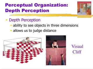
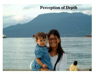
This ability to determine distance is called depth perception
The amount of light that enters the eye through the pupil is proportional to the area of the pupil or to the square of the diameter of the pupil. • The pupil of the human eye can become as small as about 1.5 millimeters and as large as 8 millimeters in diameter. • The quantity of light entering the eye can change about 30-fold as a result of changes in pupillary aperture.
“Depth of Focus” of the Lens System Increases with Decreasing Pupillary Diameter. • The greatest possible depth of focus occurs when the pupil is extremely small. • The reason for this is that, with a very small aperture, almost all the rays pass through the center of the lens, and the central-most rays are always in focus
A person normally perceives distance by three main means: • (1) the sizes of the images of known objects on the retina • (2) the phenomenon of moving parallax, • (3) the phenomenon of stereopsis.
Determination of Distance by Sizes of Retinal Images of Known Objects. • If one knows that a person being viewed is 6 feet tall, • one can determine how far away the person is simply by the size of the person’s image on the retina. • One does not consciously think about the size, but the brain has learned to calculate automatically from image sizes the distances of objects when the dimensions are known.
Determination of Distance by Moving Parallax. If a person looks off into the distance with the eyes completely still, he or she perceives no moving parallax, but when the person moves his or her head to one side or the other, the images of close-by objects move rapidly across the retinas, while the images of distant objects remain almost completely stationary.
Determination of Distance by Moving Parallax. Thus, by using this mechanism of moving parallax, one can tell the relative distances of different objects even though only one eye is used.
Determination of Distance by Stereopsis—Binocular Vision Because one eye is a little more than 2 inches to one side of the other eye, the images on the two retinas are different from each other. This gives a type of parallax that is present all the time when both eyes are being used. Binocular parallax Stereopsis cannot perceive distance beyond 50 to 200 feet.
Aqueous Humor Formed by the cilliary bodies into the posterior chamber (between the iris and the lens). 2-3 microliters/ min The aqueous humor is a freely flowing fluid The balance between formation and reabsorption of aqueous humor regulates the total volume and pressure of the intraocular fluid.
Formation of Aqueous Humor Active secretion by the epithelium of the ciliary processes. Secretion begins with active transport of sodium ions. Chloride and bicarbonate ions move along with them to maintain electrical neutrality. This causes osmosis of water from the blood capillaries Several nutrients like amino acids, ascorbic acid, and glucose are also transported across the epithelium by active transport or facilitated diffusion.
Out flow of Aqueous Humor through the pupil into the anterior chamber of the eye into the angle between the cornea and the iris through a meshwork of trabeculae canal of Schlemm extraocular veins
The canal of Schlemm • Thin-walled vein • Its endothelial membrane is so porous that even large protein molecules, as well as small particulate matter up to the size of red blood cells, can pass • It that it is filled only with aqueous humor rather than with blood • Aqueous veins.
Vitreous Humor Vitreous Humor is the clear gelatinous fluid in the posterior chamber (back 2/3) of the eye. The outer surface of the Vitreous humor is attached to the retina. Vitreous body, is a gelatinous mass held together by a fine fibrillar network composed primarily of greatly elongated proteoglycan molecules. Both water and dissolved substances can diffuse slowly in the vitreous humor, but there is little flow of fluid.
Intraocular Pressure • The average normal intraocular pressure is about 15 mm Hg • 12 to 20 mm Hg • Tonometry
Glaucoma IOP: > 60-70 mmHg • Cataract Cloudy and opaque area in lens Protein denaturation Coagulation