Download
1 / 43
470 likes | 1.46k Vues
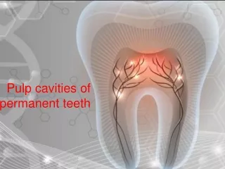
Pulp cavities of permanent teeth. Tooth structure. Tooth consist of 2 parts: crown root Crown: is the visible part Root: the part that is embedded in the jaw (not visible). Tooth structure. Enamel: The hardest, white outer part of the tooth. dentin.

E N D
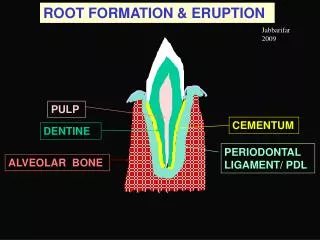
Tooth structure Tooth consist of 2 parts: crown root Crown: is the visible part Root: the part that is embedded in the jaw(not visible)
Tooth structure Enamel: The hardest, white outer part of the tooth
dentin Dentin: A layer underlying the enamel. It is a hard tissue Forms most of tooth structure
Tooth structure Cementum: A layer of connective tissue that binds the roots of the teeth firmly to the gums and jawbone pulp
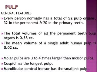
Dental pulp is a loose connective tissue has a soft, gelatinous consistency It is like heart of tooth
Nutritive: transportation of nutrient into dentin Sensory: mediation of pain sensation Defensive/reparative: formation of reparative dentin in response to irritation Formative: formation of dentin Function of pulp
Where is pulp found Pulp cavity: It is the central space in the dentin contain the dental pulp and housed it Enclosed entirely by dentin except at apical foramen
Pulp cavity Divided into: Coronal pulp chamber Pulp horn Radicular (pulp canal)
Pulp chamber Is that portion of pulp cavity located in crown
Pulp chamber Projections extend from the corners of pulp chamberinto cusps
Roof: consist of dentin covering the chamber occlusaly Pulp horn: projection of the roof under a cusp or developmental groove Floor: consist of dentin parallel to roof Canal orifice Pulp chamber
Pulp cavity Outline of pulp chamber correspond to shape of crown which it is housed Outline of pulp canal correspond to shape of roots
Radicular pulp Pulp space: Portion of pulp cavity from canal orifice to apical foramen
Type I: One canal extending from pulp chamber to the apex
Type II: Two canals arise from pulp chamber and joining into one short of the apex
Type III Two separate canals from orifice to apex , exit the root as separate foramina
Type IV One canal leaving the pulp chamber and divides into two separate canals at apical third with separate apical foramina
It is the constricted opening near the apex of the root through which the blood supply and nerves pass Apical foramen
Is a lateral branch of root canal Lateral canal
Delta system Complex system formed by breaking up of the root canal into multiple tiny canals
Pulp chamber wider mesiodistally than buccolingually Central incisor has three pulp horns corresponding three mamelones has wider pulp cavity than lateral Pulp chamber cross section is triangular Maxillary incisors
Mamelons protuberances which are present on the cutting edge of an incisor tooth when it first erupts through the gum.
Pulp chamber smaller than central Has 2 pulp horns conform mamelones Maxillary lateral incisor
Mandibular incisors Outline of pulp cavity conform to the crown 2 pulp horns Cross section: oval Pulp cavity of lateral larger than central Usually one canal but 2canals also found
Maxillary canine Widest pulp chamber in mouth labiolingually One pulp canal, one horn Cross section oval or triangular Labioligually: chamber pointed incisally, cervically wide till middle then narrow to the apex
Mandibular canine Similar to maxillary canine but less dimension
Maxillary first premolar Labiolingual: Wide pulp chamber Two root canals Buccal horn higher than lingual Cross section: kidney shape Mesiodistal: Canal narrow to apex
Maxillary first premolar Two pulp horns one under each cusp
Maxillary second premolar One root and one canal 2roots are possible 2canals in single root possible 2pulp horns Cross section: oval
Mandibular first premolar The pulp cavity of this tooth consists of two pulp horns each pulp horn is located within a cusp The buccal pulp horn is higher Majority one canal but 2 possible Cross section: oval, rectangular, round or triangular
Mandibular 2nd premolar Similar to 1st premolar with increased dimension 2-3pulp horns depend on number of cusps One root One canal
Maxillary 1st molar Cervical cross section is rhomboidal 3roots,each root one canal Mesiobuccal root may have 2 canals Cross section:canals triangular
Maxillary 1st molar Has 4 pulp horns, mesiolingual is the highest
Similar to first molar 2canals in mesiobuccal not common Orifices of canals much closer Maxillary 2nd molar
Cross section rectangular 5pulp horns 3 canals, sometimes 4 canals,mesial root has 2 canals Mandibular 1st molar
Mesial root has 2 canals one buccal and one lingual Distal root one large canal Mandibular 2nd molar Similar to 1st molar but has 4pulp horns Cross section: triangular Mandibular 1st molar