Download
1 / 40
410 likes | 437 Vues
Explore the central and peripheral nervous system divisions, including the parasympathetic and sympathetic branches, their functions, neurotransmitters, and organ responses. Learn about efferent pathways, ganglion neurons, and the role of different nerve systems in maintaining body balance.

E N D
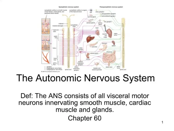
Ch. 14 The Autonomic Nervous System Parasympathetic Sympathetic
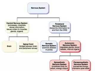
Review Central nervous system (CNS) Peripheral nervous system (PNS) Sensory (afferent) division Motor (efferent) division Somatic nervoussystem Autonomic nervous system (ANS) Sympathetic division Parasympathetic division
REVIEW … • Cranial Nerves: some with parasympathetic • Spinal Nerves: particular regions have parasympathetic and other sympathetic • Rami communicantes Ventral root Dorsal root Dorsal and ventral rootlets of spinal nerve Dorsal root ganglion Ventral ramus of spinal nerve Spinal nerve Rami communicantes
CH. 14 The Autonomic Nervous System I. Introduction– differences in Somatic Motor & Autonomic Motor • Effectors • Efferent (Motor) Pathways 1. *ANS: Number of Neurons? a) Preganglionic Neuron *- Axon is called? b) Ganglionic Neuron *- Axon is called? 2. ANS Ganglion Ganglion Ganglionic Neuron Preganglionic Neuron
I. Introduction … Slide 6 B. Efferent pathways … C. Neurotransmitters & Responses • Neurotransmitters: Which of the below nuerotransmitters for Sympathetic, Which for Parasympathetic - ANS: *Norepinephrine for: & *Acetylcholine for: • Organ (Effector) Response:
SOMATIC MOTOR Versus AUTONOMIC Somatic motor Autonomic Smooth & Cardiac Muscles Two Neurons Cell body of presynaptic neuron in Lateral Horn of Spinal cord = Preganglionic Neuron Autonomic Ganglion Cell Body of postsynaptic neuron in ganglion = Ganglionic Neuron Most axons travel in cranial & spinal nerves to effectors Neurotransmitters: Ach and NE • Skeletal Muscles • One Neuron w/ cell body in Ventral Horn of Spinal cord • No Ganglion • Axons travel in cranial and spinal nerves to muscles • Neurotransmitters: Ach
Somatic Motor vs. Autonomic Physiology Neuro- transmitter at effector Cell bodies in central nervous system Effector organs Peripheral nervous system Effect Single neuron from CNS to effector organs ACh + SOMATIC NERVOUS SYSTEM Stimulatory Heavily myelinated axon Skeletal muscle Two-neuron chain from CNS to effector organs NE ACh Unmyelinated postganglionic axon Ganglion SYMPATHETIC Lightly myelinated preganglionic axons + Epinephrine and norepinephrine ACh Stimulatory or inhibitory, depending on neuro- transmitter and receptors on effector organs AUTONOMIC NERVOUS SYSTEM Adrenal medulla Blood vessel ACh ACh Smooth muscle (e.g., in gut), glands, cardiac muscle PARASYMPATHETIC Lightly myelinated preganglionic axon Unmyelinated postganglionic axon Ganglion Acetylcholine (ACh) Norepinephrine (NE) Figure 14.2
II. ANS Divisions & Functions • Dual Innervations: Sympathetic vs. Parasympathetic • Some Organ systems have both equally • Some have one or the other Dominate • Rarely does an organ system only get input from only one
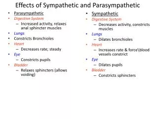
II. ANS Divisions …B. Role of the Parasympathetic DivisionPeace * Do All Below • Body maintenance: • Energy: • Organs while Relaxing • BP: • Heart: • respiratory rates: • Gastrointestinal activity: • Urinary Sys • Pupils: • Ciliary muscles:
C. Role of the Sympathetic DivisionStress * Do All Below • Fight or Flight • Mobilizes body for action or threat • BP: • Blood flow to muscles & heart: _____ • Bronchioles: __________ • Blood Glucose levels: ______ via Liver • Heart and Respiratory rates: _____________ • Digestive & Urinary Systms:
III. ANS Anatomy Eye Ciliary ganglion CN III Lacrimal gland CN VII Pterygopalatine ganglion Pterygopalatine ganglion Nasal mucosa CN IX CN X Submandibular ganglion Submandibular and sublingual glands • Overview of differences • Origin Sites • Relative Lengths of their Fibers • Location of their Ganglion • Parasympathetic Division • *Origin:preganglionic neuron cell body is in: a) *Sacral b) *Cranial • GANGLIA = Terminal (Collateral) Ganglia contains: - Locations: • Cranial & Sacral Outflow Otic ganglion Parotid gland Heart Cardiac and pulmonary plexuses Lung Liver and gallbladder Celiac plexus Stomach Pancreas S2 Large intestine S4 Pelvic splanchnic nerves Small intestine Rectum Inferior hypogastric plexus Urinary bladder and ureters Genitalia (penis, clitoris, and vagina) NEXT SLIDE
III. ANS Anatomy … B. Parasympathetic Division Eye Ciliary ganglion CN III Lacrimal gland CN VII Pterygopalatine ganglion Pterygopalatine ganglion Nasal mucosa CN IX CN X Submandibular ganglion Submandibular and sublingual glands 3. Cranial & Sacral Outflow Otic ganglion Parotid gland :Pupil & Lens Heart Midbrain Cardiac and pulmonary plexuses Lung Para for Head Pons Liver and gallbladder Celiac plexus Medulla Stomach 90% fibers Pancreas Medulla S2 (distal ½) Large intestine S4 Pelvic splanchnic nerves Small intestine Lateral Gray Horn Splanchnic n. Rectum Inferior hypogastric plexus Urinary bladder and ureters Genitalia (penis, clitoris, and vagina)
Sympathetic trunk ganglion Sympathetic trunk White ramus communicans C. Sympathetic Division Spinal cord Dorsal root • Innervates more organs • Pathway more complicated 1. Origin: *Spinal Cord? • Preganglionic Neuron *Cell Body in: • Preganglionic Axons ventral rami, then go through White Rami Communicantes - Is myelinated Ventral root Ventral ramus of spinal nerve (a) Location of the sympathetic trunk
Sympathetic trunk ganglion Sympathetic trunk White ramus communicans C. Sympathetic Division … Spinal cord Dorsal root 3.GANGLIA=Sympathetic Trunk Ganglia (Chain or Paravertebral Ganglia) • *Number: • =Are clusters of neurons linked by axonal bridges • *Contain Cell Body of ? Ventral root Ventral ramus of spinal nerve (a) Location of the sympathetic trunk
23 paired paravertebral ganglia 3. GANGLIA = Sympathetic Trunk Ganglia … Eye Lacrimal gland Nasal mucosa Pons Sympathetic trunk (chain) ganglia Blood vessels; skin (arrector pili muscles and sweat glands) • Levels:3 C, 12T, 4L, 4S • Cervical & Sacral Ganglia do not directly connect to spinal cord, BUT ascend & descend via bridges Superior cervical ganglion Salivary glands Middle cervical ganglion Heart Inferior cervical ganglion Cardiac and pulmonary plexuses Lung T1 Greater splanchnic nerve Lesser splanchnic nerve Liver and gallbladder Celiac ganglion L2 Stomach Superior mesenteric ganglion White rami communicantes Spleen Adrenal medulla Kidney Sacral splanchnic nerves Lumbar splanchnic nerves Small intestine Inferior mesenteric ganglion Large intestine Rectum Preganglionic Postganglionic Genitalia (uterus, vagina, and penis) and urinary bladder Figure 14.6
i Synapse at the same level ii Synapse at a higher or lower level C. Sympathetic Division … 4. Once preganglionic axon is in trunk ganglia, one of the following occurs: a or b • Synapse in Chain Ganglia with ganglionic neuron • Then: postganglionic axon travels out the Gray Rami same level ventral rami/spinal nerve organ
Gray ramus communicans 4. Once preganglionic axon is in trunk … Examples:Arm & Legs--skin: Sweat glands, Arrector pili muscles; smooth muscle in Blood Vessels, Heart, Lungs, Lateral horn (visceral motor zone) Ventral ramus of spinal nerve Ventral root Skin (arrector pili muscles and sweat glands) White ramus communicans Sympathetic trunk ganglion To effector 1 Synapse at the same level Blood vessels Figure 14.5b (1 of 3)
4. Once preganglionic axon is in trunk … Examples … Eye Lacrimal gland Nasal mucosa Pons Sympathetic trunk (chain) ganglia Blood vessels; skin (arrector pili muscles and sweat glands) Superior cervical ganglion Salivary glands Middle cervical ganglion Heart Inferior cervical ganglion Cardiac and pulmonary plexuses Lung T1 Greater splanchnic nerve Lesser splanchnic nerve Liver and gallbladder Celiac ganglion L2 Stomach Superior mesenteric ganglion White rami communicantes Spleen Adrenal medulla Kidney Sacral splanchnic nerves 2 Lumbar splanchnic nerves Small intestine Inferior mesenteric ganglion Large intestine Rectum Preganglionic Postganglionic Genitalia (uterus, vagina, and penis) and urinary bladder Figure 14.5b (2 of 3)
Dorsal Root Ganglion • Pass through trunk without synapsing or going out the gray rami. • Where: T5 – L2 • Organs served: abdominal organs • GANGLIA = Collateral Ganglia • Location: Anterior to chain • Serve: abdominal organs - Splanchnic Nerves: Trunk Ganglion Collateral Ganglion Splanchnic Nerve T1 Greater splanchnic nerve Lesser splanchnic nerve Liver and gallbladder Celiac ganglion L2 Stomach Superior mesenteric ganglion White rami communicantes Spleen Adrenal medulla Kidney Sacral splanchnic nerves 2 Lumbar splanchnic nerves Small intestine Inferior mesenteric ganglion Large intestine Rectum Preganglionic Postganglionic Genitalia (uterus, vagina, and penis) and urinary bladder
T1 Greater splanchnic nerve Lesser splanchnic nerve Liver and gallbladder Celiac ganglion b)…ii) Splanchnic Nerves … • Collateral Ganglia … ii) Go Directly to Adrenal Gland: • synapse with neurons in Medulla of Adrenal Gland hormones • Hormones released are the same as the neurotransmitters of same name– Epinephrine & Norepinephrine L2 Stomach Superior mesenteric ganglion White rami communicantes Spleen Adrenal medulla Kidney Sacral splanchnic nerves 2 Lumbar splanchnic nerves Small intestine Inferior mesenteric ganglion Large intestine Rectum Preganglionic Postganglionic Genitalia (uterus, vagina, and penis) and urinary bladder Adrenal medulla Hormones ACh
i • Broad Influence of preganglionic neurons: on diverse array of organs • Each Preganglionic axon branches: 10-20 • Pathways: all above possible (see 4-a & b) • Result: Diverse organs targeted to act together - Function: better emergency response • Example: As Response to Threat Heart and breathing rate increase, blood flow to skeletal muscles increases, blood flow to digestive organs decreases, and sweat gland output increases, … T1 L2
Parasympathetic vs. Sympathetic: structural differences Parasympathetic Sympathetic Location: Thoracolumbar Preganglionic Axon: short Travels from lateral horn Ventral root --> white rami Trunk Ganglia. THEN: 1) Synapse gray rami c. Ventral rami/Spinal Nerve peripheral nerve organ 2) Splanchnic nerves Collateral Ganglia & synapse . organs OR pass thru to Adrenal G. Postganglionic Axon: long Ganglia: Trunk Ganglia close to the spinal cord and Collateral Ganglia anterior to and farther away from the spinal cord • Location: Craniosacral • Preganglionic Axon: long • Travels from 1)lateral horn ventral root ventral rami nerves Terminal Ganglia Organ OR 2)brain nerve Terminal Ganglia Organ • Postganglionic Axon: short • Ganglia: Terminal Ganglia in or near effectors
ANS anatomy-Differences Parasympathetic Sympathetic Eye Eye Brain stem Salivary glands Skin* Cranial Salivary glands Sympathetic ganglia Heart Cervical Lungs Lungs T1 Heart Stomach Thoracic Stomach Pancreas Liver and gall- bladder Pancreas L1 Adrenal gland Liver and gall- bladder Lumbar Bladder Bladder Genitals Genitals Sacral Figure 14.3
IV. Physiology A. Interactions of the Autonomic Divisions • Dynamic Antagonism: Most organs have both Symp & Parasymp controlling them FUNCTION: precise control of visceral activity & Homeostasis • Autonomic Tone: When Para or Sym normally Dominates certain organs • Sympathetic Tone • Sympathetic controls: blood vessels blood pressure
2-a … Sympathetic Tone … = vasomotor tone • Normal state of Blood vessels: are always partially constricted • Exception: skeletal muscles blood vessels dilated • Parasympathetic Tone • Parasympathetic division dominates: heart, digestive and urinary tract organs • Sympathetic division overrides: during Stress
3. Cooperative Effects Parasympathetic Sympathetic Eye Eye SEX Brain stem Salivary glands Skin* • Parasympathetic fibers cause vasodilation = erection of penis or clitoris • Sympathetic fibers = ejaculation in males, contraction of vagina Cranial Salivary glands Sympathetic ganglia Heart Cervical Lungs Lungs T1 Heart Stomach Thoracic Stomach Pancreas Liver and gall- bladder Pancreas L1 Adrenal gland Liver and gall- bladder Lumbar Bladder Bladder Genitals Genitals Sacral
4. Unique Roles of Sympathetic Division • Adrenal medulla, sweat glands, arrestor pili muscles, kidneys, and most blood vessels receive only sympathetic fibers • The sympathetic division controls factors needed to Respond to STRESS: • Heat regulation: • Kidneys and B.P. to: Release of renin from kidneys to increase B.P. • Metabolic effects: • metabolic rates of cells • blood glucose levels • Mobilizes fats for fuel • Skeletal muscles contract: due to Adrenal hormones
5. Localized Versus Diffuse Effects • Parasympathetic division: short-lived, highly localized control over effectors • Sympathetic division: long-lasting, body-wide effects (NE and E breakdown slowly in liver) Two-neuron chain from CNS to effector organs NE ACh Unmyelinated postganglionic axon Ganglion SYMPATHETIC Lightly myelinated preganglionic axons + Epinephrine and norepinephrine ACh Stimulatory or inhibitory, depending on neuro- transmitter and receptors on effector organs AUTONOMIC NERVOUS SYSTEM Adrenal medulla Blood vessel ACh ACh Smooth muscle (e.g., in gut), glands, cardiac muscle PARASYMPATHETIC Lightly myelinated preganglionic axon Unmyelinated postganglionic axon Ganglion
B. Visceral Reflexes • Similar to Somatic Reflexes 1. Visceral Sensory function: • Sensory information: chemical changes, stretch, and irritation • Cell Body location: Dorsal Root Ganglia & Cranial Nerve Ganglia 2. Integration center: Interneurons in spinal cord or brain 3. Visceral Motor Neuron– preganglionic neuron: Lateral Horn of Spinal Cord • Sympathetic:Axon synapses in Chain Ganglia ganglionic, then neuron’s axon Autonomic nerves (splanichic) or spinal nerves organ/glands
Visceral Reflexes Stimulus Dorsal root ganglion Sensory receptor in viscera 1 Spinal cord Visceral sensory neuron 2 Integration center • May be preganglionic neuron (as shown) • May be a dorsal horn interneuron • May be within walls of gastrointestinal tract 3 Autonomic ganglion Efferent pathway (two-neuron chain) • Preganglionic neuron • Ganglionic neuron 4 Visceral effector 5 Response Figure 14.7
2. Parasympathetic: Axon synapses in terminal ganglia ganglionic neuron’s axon organ • Examples: emptying bladder and rectum • Example: Baroreceptors in Carotid Artery and Aortic Arch detect stretch when blood pressure or volume increase glossopharyngeal & vagus nerves Integration Center is in the Medulla Preganglionic Neuron in Medulla vagus nerve terminal ganglion with ganglionic neuron (Inhibition of sympathetic affect axon to spinal cord Trunk Ganglia with ganlionic neuron axon spinal nerve blood vessels to cause)
Visceral pain afferents travel same pathway as somatic pain fibers Pain stimuli in viscera perceived as somatic C. Referred Pain Heart Lungs and diaphragm Liver Heart Gallbladder Liver Appendix Stomach Pancreas Small intestine Ovaries Colon Kidneys Urinary bladder Ureters Figure 14.8
V. Control of ANS Functioning Communication at subconscious level Cerebral cortex (frontal lobe) Limbic system (emotional input) Hypothalamus Overall integration of ANS, the boss Brain stem (reticular formation, etc.) Regulation of pupil size, respiration, heart, blood pressure, swallowing, etc. Spinal cord Urination, defecation, erection, and ejaculation reflexes Figure 14.9
A. Hypothalamus • Overall Functions: Major controller of Autonomic & Endocrine activities • Integrates inputs: top of heirarchy 2. Centers for: heart, blood pressure, body temp, water balance, emotional states, biological drives 3. Certain Output is direct to and by way of lower brain regions, especially the nuclei of the Reticular Formation (in brain stem) • Interacts with other Brain Regions • Medulla • Heart • Respiration • Gastrointestinal Activities • Pons
C. Pons: Respiration D. Midbrain: Pupil diameter and lens focus E. Cerebral Cortex: Emotional Aspects • Unconscious modification via Limbic Association Region • Spinal Cord Level: Defecation & micturition, erection, and ejaculation • which are then affected by conscious cerebral cortex
END • Review Questions Follow
Review Questions Body maintenance is regulated by the _______________ division and its nerves arise from the ____________ regions. Mobilization is regulated by the ___________ division and its nerves arise from the _____________ region parasympathetic craniosacral sympathetic thoracolumabar
Review Questions Sympathetic fibers that control sweat glands and arrector pili muscles synapse in the ______________ _______ ganglia. Pathways that serve the intestines and liver synapse in ____________ ganglia. All preganglionic ANS figers release ____________. Sympathetic post ganglionic fibers tend to release _______________. sympathetic trunk collateral Acetylcholine (ACh) norepinephrine (NE)
Review Questions Most blood vessel smooth muscle and arrector pili are only innervated by _____________ fibers. What are two other structures that only receive these fibers? Which region of the brain is king of the ANS? How is it linked to conscious awareness? sympathetic Kidney, sweat glands, adrenal medulla Hypothalamus Limbic System
Somatic Motor vs. Autonomic Physiology Neuro- transmitter at effector Cell bodies in central nervous system Effector organs Peripheral nervous system Effect Single neuron from CNS to effector organs ACh + SOMATIC NERVOUS SYSTEM Stimulatory Heavily myelinated axon Skeletal muscle Two-neuron chain from CNS to effector organs NE ACh Unmyelinated postganglionic axon Ganglion SYMPATHETIC Lightly myelinated preganglionic axons + Epinephrine and norepinephrine ACh Stimulatory or inhibitory, depending on neuro- transmitter and receptors on effector organs AUTONOMIC NERVOUS SYSTEM Adrenal medulla Blood vessel ACh ACh Smooth muscle (e.g., in gut), glands, cardiac muscle PARASYMPATHETIC Lightly myelinated preganglionic axon Unmyelinated postganglionic axon Ganglion Acetylcholine (ACh) Norepinephrine (NE) Figure 14.2