ORBIT
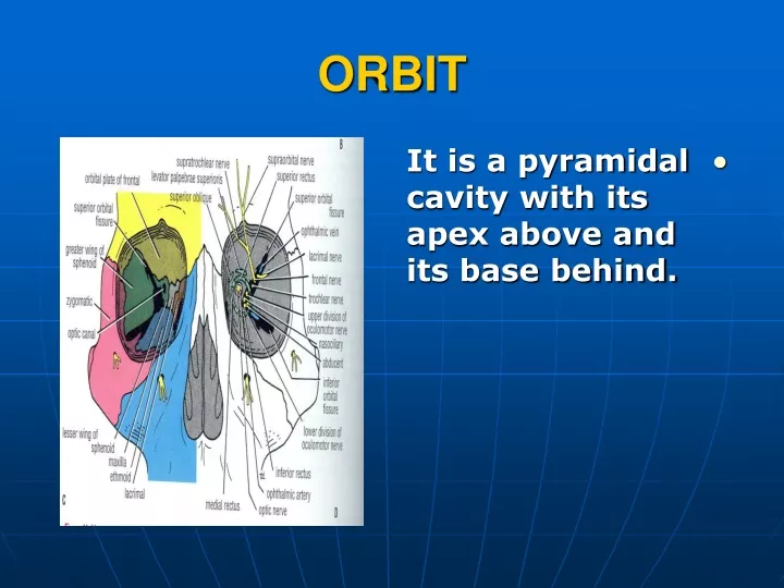
ORBIT. It is a pyramidal cavity with its apex above and its base behind. CONTENTS. (1) Eye ball . (2) Extraocular muscles. (3) Bulbar fascia. (4) Nerves . (5) Vessels . (6) Orbital fat. ORBITAL FASCIA. It is the periosteum covering the bones of the walls of the orbit.

ORBIT
E N D
Presentation Transcript
ORBIT • It is a pyramidal cavity with its apex above and its base behind.
CONTENTS • (1) Eye ball. • (2) Extraocular muscles. • (3) Bulbar fascia. • (4) Nerves. • (5) Vessels. • (6) Orbital fat.
ORBITAL FASCIA • It is the periosteum covering the bones of the walls of the orbit. • It is continuous through the foramina and fissures with the periosteum on the outer surface of the bones.
ORBITAL FASCIA • In case of : • Superior orbital fissure. • Optic canal. • Anterior ethmoidal canal, it is continuous with the endosteal layer of the dura.
ORBITAL FASCIA • The inferior orbitalfissure is bridged by the muscle of Muller (orbitalis) muscle.
EXTRAOCULAR MUSCLES • A. Levator palpebrae superioris. • B. Four recti(superior, inferior, medial andlateral). • C. Two oblique(superior and inferior).
LEVATOR PALPEBRAE SUPERIORIS • Origin : • Undersurface of the lesser wing of the sphenoid above the optic canal.
LEVATOR PALPEBRAE SUPERIORIS • The muscle ends anteriorly in a wideaponeurosis. • This aponeurosis splits into twolamellae : • Superior and Inferior.
LEVATOR PALPEBRAE SUPERIORIS • Insertion : • Suprior lamella : • To the superior tarsalplate and the skin of the upper eyelid. • Inferior lamella : • Contains(Muller’smuscle) • It isinserted into the superior tarsal plate.
LEVATOR PALPEBRAE SUPERIORIS • Nerve supply : • Oculomotor nerve (superior division). • Muller’ muscle :superior cervical sympathetic ganglion.
LEVATOR PALPEBRAE SUPERIORIS • Action : • Elevation of the upper eye lid. • Sympathetic stimulation causes further elevation of the eye lid.
PTOSIS • It is Dropping of the upper eye lid. • It is due to division of the oculomotor nerve or the cervical sympathetic ganglion (Horner’s syndrome).
THE RECTI • Origin : • From the common tendinousring (it is thickened periosteum which surrounds the optic canal and bridges over the medial end of the superior orbital fissure).
THE RECTI • The muscles are attached to the ring in positions implied by their names. • The lateral rectusarises by two heads.
THE RECTI • Insertion : • The muscles pass forwards to the sclera to be inserted (6mm) behind the cornea(in front the equator of the eyeball).
THE RECTI • Nerve supply : • 1. Oculomotornerve : to Superior, Inferior and Medial recti. • 2. Abducent nerve: toLateral rectus.
THE RECTI • Action : • The medialrectus rotates the corneamedially. • The lateral rectus rotates the cornealaterally.
THE RECTI • Action : • Superiorand Inferior recti elevateand depressthe cornea respectively (in the transverse axis).
THE RECTI • The superior and inferior recti are inserted medial to the vertical axis of the eye ball. • So they can rotate the cornea medially.
SUPERIOR OBLIQUE • Origin : • Body of the sphenoid above the common tendinous ring. • It hooks around a fibrocartilaginous pulley (trochlea) on the superomedial border of the front of the orbit.
SUPERIOR OBLIQUE • Insertion : • Into the sclera beneath the superior rectus and behind the coronalequator of the eye ball. • Nerve supply : • Trochlear nerve.
INFERIOR OBLIQUE • Origin : • Anteromedial floor of the orbit. • Insertion : • Posterolateral part of the sclera behind the equator. • Nerve supply :oculomotor nerve (inferior division).
ACTION • Superior oblique: • Rotates the corneadownwards. • Inferior oblique: • Rotates the corneaupwards. • Both muscles rotate the cornealaterally.
ACTION • The line of pull of both muscles pass medial to the vertical axis and behind the equator.
FASCIAL SHEATH OF THE EYEBALL • It separates the eye ball from the orbital fat and forms a socket for its free movement. • It surrounds the eyeball from the optic nerve to the corneoscleral junction.
FASCIAL SHEATH OF THE EYEBALL • It is pierced by the tendons of the orbital muscles. • It is reflected onto each of them as a tubular sheath.
FASCIAL SHEATH OF THE EYEBALL • Thickening of the fascia is attached to the lacrimal and zygomatic bones. • It forms the medial and lateralcheck ligaments. • Inferiorly, it forms the suspensory ligament which connects the check ligaments. • The eyeball is suspended from the medial and lateral walls of the orbit.
LACRIMAL APPARATUS • It is composed of the structures producing lacrimal fluid and controlling its passage to the nasal cavity. • They are : • Lacrimal gland and its ducts. • Conjunctival sac. • Lacrimal sac. • Naso lacrimal duct.
LACRIMAL GLAND • It is a serous gland almond in shape in the supero lateral angle of the orbit behind the upper lid. • It consists of a large orbital part and a small palpebral part.
LACRIMAL GLAND • The two parts are continuous at the lateral margin of the aponeurosis of levator palpebrae superioris. The gland has (6- 12) ducts that open into the lateral part of the superior fornix.
LACRIMAL GLAND • Nerve supply: • Facial nerve(through greater petrosal and nerve of pterygoid canal ) to the pterygopalatine ganglion. • The post ganglionic fibers pass to the gland through the zygomatic branch of zygomaticotemporal or through the lacrimal nerve.
LACRIMAL CANALICULUS • Tears accumulate in the lacus lacrimalisbefore it enters the lacrimal punctuae . • LACRIMAL CANALICULUS • They are two. Each about (10) mm long. They pass from the lacrimalpunctum in each eye lid to the lacrimal sac.
LACRIMAL SAC • It is a thin fibrous sac on the medial side of the orbit in the lacrimal fossa. It receives both canaliculi and drains to the nasolacrimal duct.
NASO LACRIMAL DUCT • It descends in the medial wall of the orbit. • It opens into the inferior meatus of the nose. Its opening is guarded by a flap of mucous membrane which prevents air passing up the duct when the nose is blown.
LACRIMAL APPARATUS • The lacrimal fluid constantly washes the front of the eye ball and its conjunctival covering . • It is drained by plinking when the increased intra conjunctival pressure produced by the closed eye lids forces the fluid into the lacrimal puncta.
CONCLUSION OF ACTION • (1) R. Superior rectus + L. inferior oblique. • (2) Superior rectus + inferior oblique (both eyes). • (3) R. Inferior oblique + L. Superior rectus.
CONCLUSION OF ACTION • (4) R. Lateral rectus + L. Medial rectus. • (5) Fixed primary position. • (6) R. Medial rectus + L. Lateral rectus.
CONCLUSION OF ACTION • (7) R. Inferior rectus + L. Superior oblique. • (8) Inferior recti + Superior oblique of both eyes. • (9) R. Superior oblique + L. Inferior rectus.