Download
1 / 22
220 likes | 348 Vues
Concerted action of poly(A) nucleases and decapping enzyme in mammalian mRNA turnover. Akio Yamashita 1, 2 , Tsung -Cheng Chang 1, 2 , Yukiko Yamashita 1 , Wenmiao Zhu 1 , Zhenping Zhong 1 , Chyi -Ying A Chen 1 & Ann-Bin Shyu. Goals of the paper.

E N D
Concerted action of poly(A) nucleases and decapping enzyme in mammalian mRNA turnover Akio Yamashita1, 2, Tsung-Cheng Chang1, 2, Yukiko Yamashita1, Wenmiao Zhu1, Zhenping Zhong1, Chyi-Ying A Chen1 & Ann-Bin Shyu
Goals of the paper • Do these deadenylases exist and act as they do in yeast turnover of mRNA • How do they affect the kinetics of normal and NMD mRNA turnover • What effect does decapping have on these processes
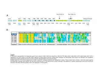
Analogs to yeast cytoplasmic deadenylation factors are found in mammalian cells Figure 1a Endogenous protein localization in NIH3T3 cells based on western blot analysis of sub cellular distributions on the (Left) Transfected HA-tagged CCR4a and CCR4b distribution analysis by western blot (Top Right) (Bottom Right) mRNA knockdown of endogenous CCR4a and b Figure 2a Confocal microscopy of tagged proteins in live cells further confirm the western blot data Cytoplasm (predominantly): CCR4, CAF, PAN2 and PAN3 Nucleus: PARN
These proteins form similar complexes to their yeast analogs Figure 1b Protein interactions were detected by co-IP in Figure 1b using HA tagged proteins. CCR4a/b-CAF1a PAN2-PAN3
Deadenylation proteins shuttle between the nucleus and cytoplasm Leptomycin B: inhibits exportin-1 Figure 2b Nuclear localization of deadenylation complexes when nuclear export is blocked Figure 2c PARN is found in the nucleus of cyclohexamide fused HeLa cells
Biphasic deadenylation of mRNAs Figure 3 Deadenlyation occurs in 2 distinct steps: shortening of the poly a tail (synchronous); further shortening of the tail and degradation of the mRNA body (asynchronous) Phase 2 appears sooner in PTC containing mRNAs
Pan2 and Ccr4 act sequentially in biphasic deadenylation (Figure 4) CCR2 mediates phase 2 degradation and PAN2 mediates phase 1 of deadenylation of b-globin mRNA as determined by overexpression. PARN does not affect the course of this mRNA despite higher expression levels Inactive variants of the proteins are transfected to verify that it is not an artifact of overexpression
Because PARN does something Figure 4 In vitro degradation of Poly(A) substrate by WT is greater than that of MT however it is not a complete loss of function
Reduction of phase 2 activity by CCR4 RNAi Figure 5 siRNA knockdown of CCR4a/b prevents the onset of asynchronous degradation PARN has supposedly very little effect on the kinetics of decay
Nucleases in NMD Figure 3 Control experiment for normal NIH3T3 cells Figure 6 Over expression of the above labeled constructs in the NIH3T3 cells compared to control results from figure 3 NMD Ccr4 and Pan2 play roles identical to that of normal mRNA processing in NMD
Nucleases in NMD cont. Figure 6 NMD under condition of reduced CCR4 and PARN expression by siRNA knockdown As before, knockdown of CCR4a/b stabilizes the mRNA
Deadenylation of mRNA can occur independently of decapping in eukaryotes Accumulation of intermediates can be detected in the absence of decapping Dcp2 indicating that Dcp2 likely acts after the first phase has completed This also seems to indicate that some mRNA must be decapped prior to deadenylation by Ccr4
Deadenylation can occur independently of decapping in eukaryotes cont. Moderate stabilization of PTC mRNA may indicate mammals depend on decapping mediated decay compared to yeast, which is almost entirely independent of decapping.
Conclusions • PAN2 is primarily involved with the initial degradation of the poly(A) tail • CCR4 is responsible for asynchronous degradation of the mRNA • Both are functionally linked and involved similarly in NMD
Questions • What are the factors involved in the interaction between deadenylation and decapping • How do these factors respond to different mRNA substrates • How does altering shuttling activity of the protein affect function in deadenylation
Decapping and Decay of Messenger RNA Occur in Cytoplasmic Processing Bodies UjwalSheth and Roy Parker
Claims • mRNA decapping and 5’-3’ degradation occurs in discrete cytoplasmic foci (P bodies) • Proteins that activate or catalyze decapping are found in P bodies • Inhibiting mRNA turnover prior to decapping leads to reduced P bodies; however, inhibiting turnover at or after decapping increases the number of P bodies • Degradation mediates are localized to P bodies
Many proteins involved in mRNA turnover localize to punctate structures Figure 1 (Left) Dcp1, Dcp2, Pat1, Lsm1,Ccr4 (discrete, transient), Dhh1 and Xrn1 with fluorescent epitopes have a punctate distribution in cells Figure 2 Mutations of the mentioned proteins and a look at distribution with P body marker Dhh1 GFP tagged Dcp1 and Xrn1 mutants show increased P body size and number while Ccr4 (deadenylation) and Pat1(Decapping activator) mutants show marked reduction
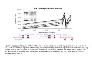
Degradation mediates are localized to P bodies Figure 3a Poly G inserted in MFA2to trap the decay intermediate; therefore, no accumulation when not present Figure 3b Co-localization of these intermediates with Lsm1 indicating that mRNAs in the process of decay are associated with P bodies Figure 3c Reproducible results using the PGK reporter mRNA
Questions • What factors are required for the localization of mRNA in P bodies? Pat1 vs Lsm1 • Are some of these factors only transiently associated with P bodies and what would that time course be? • Are P bodies associated with NMD