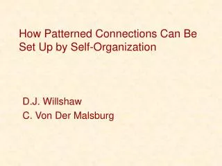
How Patterned Connections Can Be Set Up by Self-Organization
180 likes | 376 Vues
How Patterned Connections Can Be Set Up by Self-Organization. D.J. Willshaw C. Von Der Malsburg. Early Visual Pathway. Retinal ganglion cells project to LGN of the Thalamus and optic tectum in midbrain Optic tectum is the primary visual area in lower vertebrates (e.g. frogs, fish).

How Patterned Connections Can Be Set Up by Self-Organization
E N D
Presentation Transcript
How Patterned Connections Can Be Set Up by Self-Organization D.J. Willshaw C. Von Der Malsburg
Early Visual Pathway • Retinal ganglion cells project to LGN of the Thalamus and optic tectum in midbrain • Optic tectum is the primary visual area in lower vertebrates (e.g. frogs, fish)
Outline • 2 early hypothesis for map formation • Gradient models • Correlated activity models • Willshaw and von der Malsburg’s model • Retinal waves
How are maps initially formed? 2 possibilities: • Axons project randomly. Only appropriate connections with congruent activity survive. Paul Weiss OR • Chemospecificity Hypothesis. Axons are guided to targets via chemical markers. Roger Sperry
Chemospecificity Hypothesis • Retinal axons returned to original, maladaptive tectal targets
Gradient Models • topographic branching results from repulsive ligand gradients • Growth cones have different densities of ligand receptors • Multiple ligands create complex branching
Example Ligands • Ephrin-A family • boundaries vary Monschau et al. (1997).
Q: How do maps become fine-tuned?A: Correlated neural activity tectum retina all-to-all connectivity selective connectivity Input layer neighbors output layer neighbors
Willshaw & von der Malsburg 1976 • Sperry-type models assume axons seek targets independently using neuron specific labels • W & vdM’s model uses the lateral connections within input and output layers • Goal of model is to encode the geometrical proximity of input cells using their correlated neural activity.
General Structure retina tectum • Short range excitatory connections • Long range inhibitory connections • Competitive, Hebbian synapses • Spontaneous activity within input layer
Equations Hj* = activity in post-syn cell j Ai* = state of pre-cell i; 1 if active at time t, 0 otherwise sij = connection weight i j ekj = excitatory connection of post- cell k post-cell j ikj = inhibitory connection of post- cell k post-cell j Weight update: Normalization: M = # pre cells N = # post cells
Orientation of the map • orientation of map can be fixed using polarity markers • bias weights of a small pre-syn region in the desired orienation with a small post-syn region
Mapping results • Mean coordinates of weighted pre-cells projecting to each post-cell. • Maps shift to accommodate new cells.
Correlated Firing: Retinal Waves • Segregation of retinal inputs in LGN is complete before birth • TTX on optic chiasm disrupts segregation, suggests activity dependence • Spontaneous waves of synchronous RGC firing might organize mapping Feller et al, (1996)
Properties of Retinal Waves • Occur spontaneously • Appear randomly • Spread to a limited region: local excitation; global inhibition
Summary • Retino-tectal maps are initially formed using chemical gradients. • Correlated activity is used to fine tune connections. • Exploiting lateral connections allows for more efficient genetic coding versus Sperry type models. • Retinal waves share many properties of Willshaw and von der Malsburg’s model.