Brain & Cranial Nerves
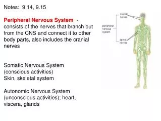
Brain & Cranial Nerves. Dr. Michael P. Gillespie. Major Parts of the Brain. Brain stem Cerebellum Diencephalon Cerebrum. Brain Stem. Continuous with the spinal cord. Subdivisions Medulla Oblongata Pons Midbrain. Cerebellum. Posterior to the brain stem. Cerebellum = little brain.


Brain & Cranial Nerves
E N D
Presentation Transcript
Brain & Cranial Nerves Dr. Michael P. Gillespie
Major Parts of the Brain • Brain stem • Cerebellum • Diencephalon • Cerebrum
Brain Stem • Continuous with the spinal cord. • Subdivisions • Medulla Oblongata • Pons • Midbrain
Cerebellum • Posterior to the brain stem. • Cerebellum = little brain.
Diencephalon • Superior to the brain stem. • Subdivisions • Thalamus • Hypothalamus • Epithalamus • Di = through; encephalon = brain
Cerebrum • Supported on the diencephalon and brain stem. • Largest part of the brain. • Cerebrum = brain.
Brain Blood Supply • Arteries • Internal carotid arteries • Vertebral arteries • Veins • Internal jugular veins
Brain Blood Flow • The brain consumes about 20% of the oxygen and glucose used at rest. • A brief slowing of blood flow may cause unconsciousness. • When activity of neurons and neuroglia in a certain portion of the brain increases, blood flow to that region increases.
Brain Blood Flow • An interruption of blood flow for 1 to 2 minutes impairs neural function. • Total deprivation of oxygen for 4 minutes causes permanent injury. • If the blood entering the brain has a low level of glucose, mental confusion, dizziness, convulsions, and loss of consciousness may occur.
Blood Brain Barrier • The blood-brain barrier (BBB) protects the brain from harmful substances and pathogens. • It prevents the passage of many substances from the blood to the brain tissue. • Tight junctions seal together endothelial cells of brain capillaries. • Astrocytes selectively allow some substances through and not others.
Permeability of the BBB • Water-soluble substances. • Glucose crosses the BBB by active transport. • Creatinine, urea, and most ions cross the BBB very slowly. • Proteins and most antibiotic drugs do not cross the BBB. • Lipid-soluble substances. • Oxygen, carbon dioxide, alcohol, most anesthetic agents cross easily.
Breaching the BBB • The BBB prevents the passage of harmful substances into the brain, but it also prevents the passage of useful drugs. • Drugs are injected in a concentrated sugar solution to facilitate passage. • The high osmotic pressure causes cells lining the barrier to shrink and makes the membrane “leaky”.
Protective Coverings • Cranium • Meninges. • Dura mater (Outer layer). • Two dural layers around the brain and one around the spinal cord. • Arachnoid mater (Middle layer). • Pia mater (Inner layer). • No epidural space around the brain.
Protective Coverings • Extensions of dura mater separate parts of the brain. • Falx cerebri – separates the two hemispheres of the cerebrum. • Falx cerebelli – separates the two hemispheres of the cerebellum. • Tentorium cerebelli – separates the cerebrum from the cerebellum.
Cerebrospinal Fluid (CSF) • Clear colorless liquid. • Protects the brain and spinal cord from chemical and physical injuries. • Carries oxygen, glucose, and other needed chemicals from the blood to the neurons and neuroglia. • Circulates in the subarachnoid space (between the arachnoid mater and pia mater).
Formation of CSF in the Ventricles • CSF is formed in the ventricles. • Formed by ependymal cells that cover the choroid plexuses of the ventricles.
Formation of CSF in the Ventricles • There are 4 ventricles. • Functions of CSF. • Mechanical protection. • Shock absorption. • Buoys the brain. • Chemical protection – optimal chemical environment. • Circulation – medium of exchange for wastes and nutrients.
Hydrocephalus • Abnormalities of the brain can interfere with drainage of CSF from the ventricles and subarachnoid space. • CSF pressure increases causing hydrocephalus. • In infants this causes the fontanels to budge.
Hydrocephalus • Tumors, inflammation, developmental malformations can all cause hydrocephalus. • Pressure buildup can damage the delicate nervous tissue. • A surgeon can implant a drain line called a shunt to divert CSF. • In adults, hydrocephalus may occur after head injury, meningitis, or subarachnoid hemorrhage.
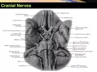
Brain Stem • Between the brain and spinal cord. • 3 regions. • Medulla oblongata. • Pons. • Midbrain.
Medulla Oblongata • A continuation of the spinal cord. • Sensory (ascending) tracts and motor (descending) tracts travel through the white matter of the medulla. • Many nerves decussate (cross over) in the medulla.
Medulla Oblongata • Cardiovascular center regulates the heartbeat and the diameter of the blood vessels.
Medulla Oblongata • The medullary rhythmicity area adjusts the rhythm of the breathing and controls reflexes for vomiting, coughing, and sneezing.
Medulla Oblongata • The nuclei for the following cranial nerves reside in the medulla: • VIII (vestibulocochlear). • IX (glossopharyngeal). • X (vagus). • XI (accessory). • XII (hypoglossal).
Pons • Pneumotaxic area and apneustic area regulate breathing. • Nuclei for cranial nerves V (trigeminal), VI (abducens), VII (facial), and VIII (vestibulocochlear).
Midbrain • The midbrain or mesencephalon contains the superior colliculi (visual actvities) and inferior colliculi (auditory pathways). • The midbrain contains the substantia nigra which release dopamine to help control subconscious muscle activities. Loss of these neurons results in Parkinson disease. • Cranial nerves III (oculomotor) and IV (trochlear) originate here.
Cerebellum • The second largest part of the brain. • A main function of the cerebellum is to evaluate how well movements are being carried out and correct for discrepancies. This helps to “smooth out” movements.
Diencephelon • Epithalamus. • Contains the pineal gland which secretes melatonin. • Thalamus. • Relays sensory information to the cortex. • Provides crude perception of touch, pressure, pain, and temperature.
Diencephelon • Subthalamus. • Controls body movements. • Hypothalamus. • Controls and integrates activities of the ANS. • Regulates emotional and behavioral patterns. • Regulates cicadian rhythms. • Regulates eating and drinking behavior. • Produces hormones oxytocin and ADH.
Cerebrum • Sensory areas interpret sensory impulses. • Motor areas control muscular movement. • Association areas function in emotional and intellectual processes. • Basal areas regulate gross muscle movements and regulate muscle tone. • Limbic system functions in survival behaviors.
Brain Injuries • Concussion – an abrupt, temporary loss of consciousness following a blow to the head. • Most common brain injury. • Signs – headache, drowsiness, lack of concentration, confusion, amnesia.
Brain Injuries • Contusion – bruising of the brain due to trauma and includes leakage of blood. • Signs - immediate loss of consciousness, transient cessation of respiration, decreased blood pressure.
Brain Injuries • Laceration – tear of the brain usually from a skull fracture or gunshot wound. • Rupture of large blood vessels. • Consequences – cerebral hematoma (localized pool of blood, usually clotted), edema, and increased intracranial pressure.
Cerebral Cortex Areas and Functions • Sensory areas – receive and interpret sensory information.