
Chapter 10- Amphibians
E N D
Presentation Transcript
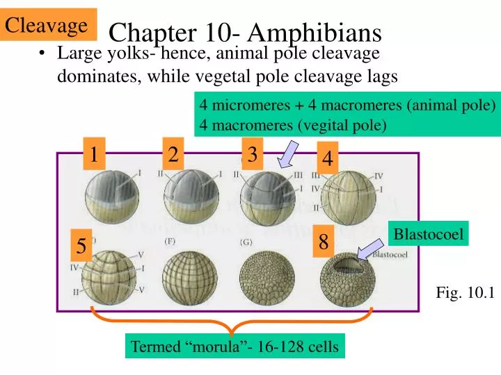
Cleavage Chapter 10- Amphibians 4 micromeres + 4 macromeres (animal pole) 4 macromeres (vegital pole) • Large yolks- hence, animal pole cleavage dominates, while vegetal pole cleavage lags 1 2 3 4 Blastocoel 8 5 Fig. 10.1 Termed “morula”- 16-128 cells
Gastrulation Use Xenopus as a model • Three tasks- • To bring inside those cells destined to be endodermal organs • To surround embryo with cells destined for ectoderm • To place cells cells “in between” destined to be mesoderm VegT establishes animal/vegital pole Ectoderm-skin/nerves Mesoderm- Blood, heart, kidney, bones Endoderm- Gut and associated organs VegT Antisense results in lack of endoderm Fig. 10.6
Gastrulation Cells migrate toward dorsal lip, then turn inward into interior Gastrulation begins in marginal zone at dorsal lip, formed by bottle cells Archenteron- formed by invading cells Blastocoel function 1. permits cell migra-tion 2. Prevents premature interactions Animal Fig. 10.7 Vegital Waves of cells continue to enter through dorsal lip Animal pole cells migrate downward and eventually encase embryo, except for yolk plug, termed epiboly (see fig. 8.6)
Gastrulation • Order of cells entry through dorsal blastopore • 1. Pharengeal endomesoderm (foregut) • 2. Notochord and somite precursors • 3. Heart, kidney and ventral mesoderm Fig. 10.7
Gastrulation Some details on Xenopus gastrulation Positioning the blastopore • The point of sperm entry marks the ventral side • The opposite side marks point of gastrulation initiation • The cortical cytoplasm rotates 30 degrees toward sperm entry point 30o Dorsal blastopore lip formation Sperm entry Fig. 7.35 90 min 0 min
How do the invading cells know to crawl along inside of animal pole? Answer- fibronectin • Cells on the blastocoel roof secrete fibronectin, which invading mesodem cells will track along • Mesoderm receptors recognize the Arg-Gly-Asp peptide • Evidence- if inject Arg-Gly-Asp peptide, disrupt involution Fig. 10.13
What dictates axis formation? Separate embryo vertically Separate embryo horizontally Answer- the Spemann organizer – aka the dorsal lip The dorsal lip is in the gray crescent Fig. 10.18
The Spemann organizer Of all tissues in early gastrula, only dorsal lip of blastopore has its cells fated Evidence Transplant dorsal lip onto an embryo, observe a second invagination and a second embryo Thus, Dorsal lip cells dictate primary embryonic induction Fig. 10.20
Yeah, but what is the mechanism of axis formation by the Spemann Organizer?? Answer- signals from vegetal cells Animal- Ectoderm Marginal- Mesoderm Vegetal-Endoderm Normal “layers” Evidence If place animal cap next to vegetal cells- animal becomes mesoderm (muscle, etc) Vegetal cells that induce the Spemann organizer are called the Nieukoop center Organizer Nieukoop center Fig. 10.21
A mechanistic model of axis formation: B-catenin accumulates in dorsal region to activate organizer genes, including goosecoid B-catenin (orange) Fig. 10.23 How does B-catenin accumulate? Hint- It is initially throughout embryo. Recall Wnt signaling pathway from ch. 6 Dishevelled expression inhibits GSK-3, thus stablezing B-catenen Fig. 6.23
A mechanistic model of axis formation: Fertilization B-catenin activates Samois expression, which activates Goosecoid expression Dishevelled Cortical rotation B-catenin protein Siamois gene Discheveled released GSK-3 inhibited TGF-b signaling Goosecoid gene Goosecoid protein B-catenin stabilized only in dorsal region Fig. 10.24
Organizer functions The ability to • become dorsal mesoderm • dorsalize surrounding ventral mesoderm into lateral mesoderm • dorsalize ectoderm into neural ectoderm • initiate gastrulation movements • cause neural plate to become neural tube
But what is goosecoid function?? - - Dishevelled Evidence of goosecoid’s importance • If increase organizer mesoderm, observe expanded region of goosecoid expression • Inject goosecoid into 4-cell ventral blastomeres- progeny blastomeres induce cell migration and form dorsal lip GSK-3 B-catenin samois • Goosecoid also recruits neighboring cells into the dorsal axis goosecoid Thus, goosecoid must activate genes involved in dorsal/ventral and anterior/posterior axes Activates two groups of proteins- Examples • Bone morphogenesis • protein (BMP) inhibitors Noggin, Chordin, Follistatin 2. Wnt inhibitors Cerberus, FRZB, DICCK
Bone morphogenesis protein (BMP) inhibitors Example- Noggin (a BMP inhibitor) How discovered?- cDNAs from a plasmid library injected into UV treated embryos. If expose embryo to ultraviolet radiation- cortical rotation fails, and embryo lacks dorsal structures Fig. 10.29 Noggin functions by binding BMP4 and BMP2, thus preventing these proteins from binding their receptors Tail + belly Normal Head + belly Chordin and follistatin also act by inhibiting BMP proteins Only head Noggin mRNA injected
2. Wnt inhibitors Example- Frzb (a Wnt inhibitor) Normal Fig. 10.35- Xwnt is inhibited by Frzb (produced by Organizer) Only head Fig. 10.36 Frzb How?? Frzb functions by binding Xwnt8, thus preventing it from binding the wnt receptor Thus, the Organizer secretes BMP and Wnt signaling inhibitors to direct head, belly and tail production Fig. 10.36
Thus, • BMP4 is the inducer of ventral ectoderm (epidermis) • BMP4 is the ventralizer of mesoderm (connective tissue, blood) • Noggin, chordin and follistatin (secreted by the Organizer) • inhibit BMP4
Regional specificity of induction The organizer not only induced the neural tube, but also specifies neural regions- forebrain, hindbrain, spinocaudel region 1st organizer cells to enter embryo induce brain and head 2nd organizer cells become spinal cord and tail Transplant young gastrula dorsal lip Two heads Transplant advanced gastrula dorsal lip Two bellies?
What molecules are involved in regional specification? • Fibroblast growth factors (FGFs)- • induces anterior neural tissue into posterior neural tissue • Induces forebrain to express hind-brain-specific genes • Retinoic Acid (RA)- • Overexpression inhibits fore and mid-brain development • as wells as prevent posterior structure formation • Induces forebrain to express hind-brain-specific genes RA 3. What molecules are involved in left-right axis formation? • Note that internal organs are not placed symmetrically- heart, gut tube • Vg1 protein activates nodalonly on left side, which activates Pitx2